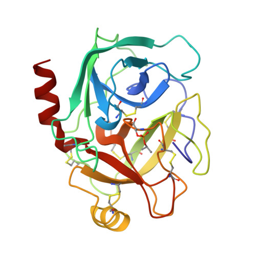
Solvent structure in crystals of trypsin determined by X-ray and neutron diffraction.
Finer-Moore, J.S., Kossiakoff, A.A., Hurley, J.H., Earnest, T., Stroud, R.M.(1992) Proteins 12: 203-222
- PubMed: 1557349
- DOI: https://doi.org/10.1002/prot.340120302
- Primary Citation of Related Structures:
5PTP - PubMed Abstract:
The solvent structure in orthorhombic crystals of bovine trypsin has been independently determined by X-ray diffraction to 1.35 A resolution and by neutron diffraction to 2.1 A resolution. A consensus model of the water molecule positions was obtained using oxygen positions identified in the electron density map determined by X-ray diffraction, which were verified by comparison to D2O-H2O difference neutron scattering density. Six of 184 water molecules in the X-ray structure, all with B-factors greater than 50 A2, were found to be spurious after comparison with neutron results. Roughly two-thirds of the water of hydration expected from thermodynamic data for proteins was localized by neutron diffraction; approximately one-half of the water of hydration was located by X-ray diffraction. Polar regions of the protein are well hydrated, and significant D2O-H2O difference density is seen for a small number of water molecules in a second shell of hydration. Hydrogen bond lengths and angles calculated from unconstrained refinement of water positions are distributed about values typically seen in small molecule structures. Solvent models found in seven other bovine trypsin and trypsinogen and rat trypsin structures determined by X-ray diffraction were compared. Internal water molecules are well conserved in all trypsin structures including anionic rat trypsin, which is 65% homologous to bovine trypsin. Of the 22 conserved waters in trypsin, 19 were also found in trypsinogen, suggesting that they are located in regions of the apoprotein that are structurally conserved in the transition to the mature protein. Seven waters were displaced upon activation of trypsinogen. Water structure at crystal contacts is not generally conserved in different crystal forms. Three groups of integral structural water molecules are highly conserved in all solvent structures, including a spline of water molecules inserted between two beta-strands, which may resemble an intermediate in the formation of beta sheets during the folding of a protein.
- Department of Biochemistry and Biophysics, University of California, San Francisco 94143-0448.
Organizational Affiliation:


