5W2H

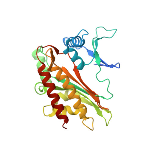
Crystal structure of the core catalytic domain of human inositol phosphate multikinase in complex with Ins(1,4,5)P3 and ADP


Experimental Data Snapshot

  • Method: X-RAY DIFFRACTION
  • Resolution: 1.90 Å
  • R-Value Free: 0.230 
  • R-Value Work: 0.163 
  • R-Value Observed: 0.167 

Starting Model: experimental
View more details

wwPDB Validation   3D Report Full Report


Ligand Structure Quality Assessment 


This is version 3.1 of the entry. See complete history


Literature

Structural features of human inositol phosphate multikinase rationalize its inositol phosphate kinase and phosphoinositide 3-kinase activities.

Wang, H.Shears, S.B.

(2017) J Biol Chem 292: 18192-18202

  • DOI: https://doi.org/10.1074/jbc.M117.801845
  • Primary Citation of Related Structures:  
    5W2G, 5W2H, 5W2I

  • PubMed Abstract: 

    Human inositol phosphate multikinase ( Hs IPMK) critically contributes to intracellular signaling through its inositol-1,4,5-trisphosphate (Ins(1,4,5)P 3 ) 3-kinase and phosphatidylinositol 4,5-bisphosphate (PtdIns(4,5)P 2 ) 3-kinase activities. This catalytic profile is not conserved; orthologs from Arabidopsis thaliana and Saccharomyces cerevisiae are predominantly Ins(1,4,5)P 3 6-kinases, and the plant enzyme cannot phosphorylate PtdIns(4,5)P 2 Therefore, crystallographic analysis of the yeast and plant enzymes, without bound inositol phosphates, do not structurally rationalize Hs IPMK activities. Here, we present 1.6-Å resolution crystal structures of Hs IPMK in complex with either Ins(1,4,5)P 3 or PtdIns(4,5)P 2 The Ins(1,4,5)P 3 headgroup of PtdIns(4,5)P 2 binds in precisely the same orientation as free Ins(1,4,5)P 3 itself, indicative of evolutionary optimization of 3-kinase activities against both substrates. We report on nucleotide binding between the separate N- and C-lobes of Hs IPMK. The N-lobe exhibits a remarkable degree of conservation with protein kinase A (root mean square deviation = 1.8 Å), indicating common ancestry. We also describe structural features unique to Hs IPMK. First, we observed a constrained, horseshoe-shaped substrate pocket, formed from an α-helix, a 3 10 helix, and a recently evolved tri-proline loop. We further found Hs IPMK activities rely on a preponderance of Gln residues, in contrast to the larger Lys and Arg residues in yeast and plant orthologs. These conclusions are supported by analyzing 14 single-site Hs IPMK mutants, some of which differentially affect 3-kinase and 6-kinase activities. Overall, we structurally rationalize phosphorylation of Ins(1,4,5)P 3 and PtdIns(4,5)P 2 by Hs IPMK.


  • Organizational Affiliation

    From the Inositol Signaling Group, Signal Transduction Laboratory, NIEHS, National Institutes of Health, Research Triangle Park, North Carolina 27709 wangh7@niehs.nih.gov.


Macromolecules
Find similar proteins by:  (by identity cutoff)  |  3D Structure
Entity ID: 1
MoleculeChains Sequence LengthOrganismDetailsImage
Inositol polyphosphate multikinase,Inositol polyphosphate multikinase257Homo sapiensMutation(s): 0 
Gene Names: IPMKIMPK
EC: 2.7.1.151 (PDB Primary Data), 2.7.1.153 (UniProt), 2.7.1.140 (UniProt)
UniProt & NIH Common Fund Data Resources
Find proteins for Q8NFU5 (Homo sapiens)
Explore Q8NFU5 
Go to UniProtKB:  Q8NFU5
PHAROS:  Q8NFU5
GTEx:  ENSG00000151151 
Entity Groups  
Sequence Clusters30% Identity50% Identity70% Identity90% Identity95% Identity100% Identity
UniProt GroupQ8NFU5
Sequence Annotations
Expand
  • Reference Sequence
Small Molecules
Ligands 4 Unique
IDChains Name / Formula / InChI Key2D Diagram3D Interactions
ADP (Subject of Investigation/LOI)
Query on ADP

Download Ideal Coordinates CCD File 
D [auth A]ADENOSINE-5'-DIPHOSPHATE
C10 H15 N5 O10 P2
XTWYTFMLZFPYCI-KQYNXXCUSA-N
I3P (Subject of Investigation/LOI)
Query on I3P

Download Ideal Coordinates CCD File 
E [auth A]D-MYO-INOSITOL-1,4,5-TRIPHOSPHATE
C6 H15 O15 P3
MMWCIQZXVOZEGG-XJTPDSDZSA-N
PO4
Query on PO4

Download Ideal Coordinates CCD File 
F [auth A]PHOSPHATE ION
O4 P
NBIIXXVUZAFLBC-UHFFFAOYSA-K
MG (Subject of Investigation/LOI)
Query on MG

Download Ideal Coordinates CCD File 
B [auth A],
C [auth A]
MAGNESIUM ION
Mg
JLVVSXFLKOJNIY-UHFFFAOYSA-N
Experimental Data & Validation

Experimental Data

  • Method: X-RAY DIFFRACTION
  • Resolution: 1.90 Å
  • R-Value Free: 0.230 
  • R-Value Work: 0.163 
  • R-Value Observed: 0.167 
  • Space Group: P 42 21 2
Unit Cell:
Length ( Å )Angle ( ˚ )
a = 78.015α = 90
b = 78.015β = 90
c = 85.915γ = 90
Software Package:
Software NamePurpose
REFMACrefinement
HKL-2000data reduction
HKL-2000data scaling

Structure Validation

View Full Validation Report


Ligand Structure Quality Assessment 


Entry History 

Deposition Data

Revision History  (Full details and data files)

  • Version 1.0: 2017-09-13
    Type: Initial release
  • Version 1.1: 2017-09-20
    Changes: Database references
  • Version 1.2: 2017-11-15
    Changes: Database references
  • Version 2.0: 2017-12-20
    Changes: Advisory, Atomic model, Database references, Derived calculations
  • Version 3.0: 2018-01-17
    Changes: Advisory, Atomic model, Database references, Derived calculations
  • Version 3.1: 2023-10-04
    Changes: Data collection, Database references, Derived calculations, Refinement description