5X0I | pdb_00005x0i

Crystal structure of PKM2 R399E mutant complexed with FBP and serine


Experimental Data Snapshot

  • Method: X-RAY DIFFRACTION
  • Resolution: 2.64 Å
  • R-Value Free: 
    0.218 (Depositor), 0.200 (DCC) 
  • R-Value Work: 
    0.180 (Depositor), 0.190 (DCC) 
  • R-Value Observed: 
    0.182 (Depositor) 

Starting Model: experimental
View more details

wwPDB Validation   3D Report Full Report


Ligand Structure Quality Assessment 

Created with Raphaël 2.3.0Worse 01 BetterLigand structure goodness of fit to experimental dataBest fitted FBPClick on this verticalbar to view details

This is version 2.1 of the entry. See complete history


Literature

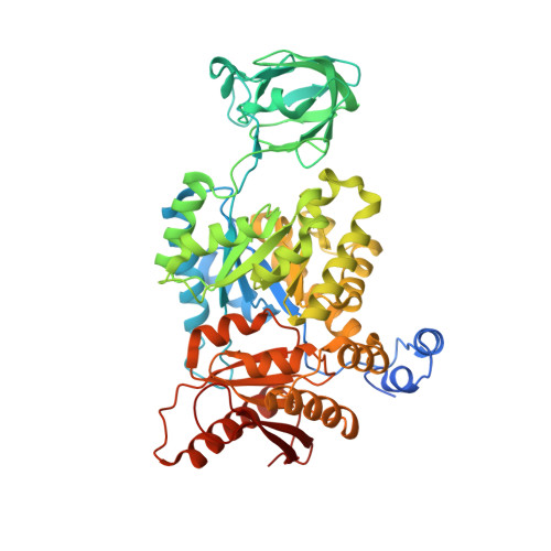
Mutations in the PKM2 exon-10 region are associated with reduced allostery and increased nuclear translocation.

Chen, T.J.Wang, H.J.Liu, J.S.Cheng, H.H.Hsu, S.C.Wu, M.C.Lu, C.H.Wu, Y.F.Wu, J.W.Liu, Y.Y.Kung, H.J.Wang, W.C.

(2019) Commun Biol 2: 105-105

  • DOI: https://doi.org/10.1038/s42003-019-0343-4
  • Primary Citation of Related Structures:  
    5X0I

  • PubMed Abstract: 

    PKM2 is a key metabolic enzyme central to glucose metabolism and energy expenditure. Multiple stimuli regulate PKM2's activity through allosteric modulation and post-translational modifications. Furthermore, PKM2 can partner with KDM8, an oncogenic demethylase and enter the nucleus to serve as a HIF1α co-activator. Yet, the mechanistic basis of the exon-10 region in allosteric regulation and nuclear translocation remains unclear. Here, we determined the crystal structures and kinetic coupling constants of exon-10 tumor-related mutants (H391Y and R399E), showing altered structural plasticity and reduced allostery. Immunoprecipitation analysis revealed increased interaction with KDM8 for H391Y, R399E, and G415R. We also found a higher degree of HIF1α-mediated transactivation activity, particularly in the presence of KDM8. Furthermore, overexpression of PKM2 mutants significantly elevated cell growth and migration. Together, PKM2 exon-10 mutations lead to structure-allostery alterations and increased nuclear functions mediated by KDM8 in breast cancer cells. Targeting the PKM2-KDM8 complex may provide a potential therapeutic intervention.


  • Organizational Affiliation

    1Institute of Molecular and Cellular Biology and Department of Life Science, National Tsing-Hua University, Hsinchu, 30013 Taiwan.


Macromolecules
Find similar proteins by:  (by identity cutoff)  |  3D Structure
Entity ID: 1
MoleculeChains Sequence LengthOrganismDetailsImage
Pyruvate kinase PKM
A, B, C, D
551Homo sapiensMutation(s): 1 
Gene Names: PKMOIP3PK2PK3PKM2
EC: 2.7.1.40 (PDB Primary Data), 2.7.11.1 (UniProt), 2.7.10.2 (UniProt)
UniProt & NIH Common Fund Data Resources
Find proteins for P14618 (Homo sapiens)
Explore P14618 
Go to UniProtKB:  P14618
PHAROS:  P14618
GTEx:  ENSG00000067225 
Entity Groups  
Sequence Clusters30% Identity50% Identity70% Identity90% Identity95% Identity100% Identity
UniProt GroupP14618
Sequence Annotations
Expand
  • Reference Sequence
Small Molecules
Ligands 5 Unique
IDChains Name / Formula / InChI Key2D Diagram3D Interactions
FBP
Query on FBP

Download Ideal Coordinates CCD File 
E [auth A],
J [auth B],
O [auth C],
T [auth D]
1,6-di-O-phosphono-beta-D-fructofuranose
C6 H14 O12 P2
RNBGYGVWRKECFJ-ARQDHWQXSA-N
SER
Query on SER

Download Ideal Coordinates CCD File 
F [auth A],
K [auth B],
P [auth C],
U [auth D]
SERINE
C3 H7 N O3
MTCFGRXMJLQNBG-REOHCLBHSA-N
PYR
Query on PYR

Download Ideal Coordinates CCD File 
G [auth A],
L [auth B],
Q [auth C],
V [auth D]
PYRUVIC ACID
C3 H4 O3
LCTONWCANYUPML-UHFFFAOYSA-N
K
Query on K

Download Ideal Coordinates CCD File 
I [auth A],
N [auth B],
S [auth C],
X [auth D]
POTASSIUM ION
K
NPYPAHLBTDXSSS-UHFFFAOYSA-N
MG
Query on MG

Download Ideal Coordinates CCD File 
H [auth A],
M [auth B],
R [auth C],
W [auth D]
MAGNESIUM ION
Mg
JLVVSXFLKOJNIY-UHFFFAOYSA-N
Experimental Data & Validation

Experimental Data

  • Method: X-RAY DIFFRACTION
  • Resolution: 2.64 Å
  • R-Value Free:  0.218 (Depositor), 0.200 (DCC) 
  • R-Value Work:  0.180 (Depositor), 0.190 (DCC) 
  • R-Value Observed: 0.182 (Depositor) 
Space Group: P 21 21 21
Unit Cell:
Length ( Å )Angle ( ˚ )
a = 116.601α = 90
b = 137.875β = 90
c = 149.709γ = 90
Software Package:
Software NamePurpose
REFMACrefinement
HKL-2000data reduction
HKL-2000data scaling
MOLREPphasing

Structure Validation

View Full Validation Report


Ligand Structure Quality Assessment 

Created with Raphaël 2.3.0Worse 01 BetterLigand structure goodness of fit to experimental dataBest fitted FBPClick on this verticalbar to view details

Entry History & Funding Information

Deposition Data


Funding OrganizationLocationGrant Number
NHRITaiwanNHRI-EX105-10517BI

Revision History  (Full details and data files)

  • Version 1.0: 2018-01-31
    Type: Initial release
  • Version 1.1: 2019-04-03
    Changes: Data collection, Database references
  • Version 1.2: 2019-11-20
    Changes: Database references
  • Version 1.3: 2020-07-29
    Type: Remediation
    Reason: Carbohydrate remediation
    Changes: Data collection, Derived calculations, Structure summary
  • Version 2.0: 2023-11-15
    Changes: Atomic model, Data collection, Database references, Derived calculations, Structure summary
  • Version 2.1: 2023-11-22
    Changes: Refinement description