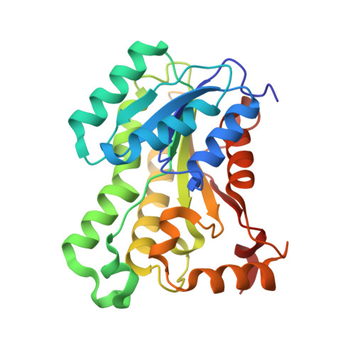
5YCS

X-Ray Structure of Enoyl-Acyl Carrier Protein Reductase from Bacillus Anthracis with triclosan


Experimental Data Snapshot

  • Method: X-RAY DIFFRACTION
  • Resolution: 1.95 Å
  • R-Value Free: 0.190 
  • R-Value Work: 0.160 
  • R-Value Observed: 0.161 

Starting Model: experimental
View more details

wwPDB Validation   3D Report Full Report


Ligand Structure Quality Assessment 


This is version 1.1 of the entry. See complete history


Literature

Structural insights into the dimer-tetramer transition of FabI from Bacillus anthracis

Kim, H.T.Kim, S.Na, B.K.Chung, J.Hwang, E.Hwang, K.Y.

(2017) Biochem Biophys Res Commun 493: 28-33

  • DOI: https://doi.org/10.1016/j.bbrc.2017.09.084
  • Primary Citation of Related Structures:  
    5YCR, 5YCS, 5YCV, 5YCX

  • PubMed Abstract: 

    Enoyl-ACP reductase (ENR, also known as FabI) has received considerable interest as an anti-bacterial target due to its essentiality in fatty acid synthesis. All the FabI structures reported to date, regardless of the organism, are composed of homo-tetramers, except for two structures: Bacillus cereus and Staphylococcus aureus FabI (bcFabI and saFabI, respectively), which have been reported as dimers. However, the reason for the existence of the dimeric form in these organisms and the biological meaning of dimeric and tetrameric forms of FabI are ambiguous. Herein, we report the high-resolution crystal structure of a dimeric form of Bacillus anthracis FabI (baFabI) and the crystal structures of tetrameric forms of baFabI in the apo state and in complex with NAD + and with NAD + -triclosan, at 1.7 Å, 1.85 Å, 1.96 Å, and 1.95 Å, respectively. Interestingly, we found that baFabI with a His 6 -tag at its C-terminus exists as a dimer, whereas untagged-baFabI exists as a tetramer. The His 6 -tag may block the dimer-tetramer transition, since baFabI has relatively short-length amino acids ( 255 LG 256 ) after the 3 10 -helix η7 compared to those of FabI of other organisms. The dimeric form of baFabI is catalytically inactive, because the α-helix α5 occupies the NADH-binding site. During the process of dimer-tetramer transition, this α5 helix rotates about 55° toward the tetramer interface and the active site is established. Therefore, tetramerization of baFabI is required for cofactor binding and catalytic activity.


  • Organizational Affiliation

    Crystalgenomics, Inc., 5F, Tower A, Korea Bio Park 700, Daewangpangyo-ro, Bundang-gu, Seongnam-si, Gyeonggi-do 13524, South Korea; Division of Biotechnology, College of Life Sciences and Biotechnology, Korea University, 145 Anam-ro, Seongbuk-gu, Seoul 02841, South Korea.


Macromolecules
Find similar proteins by:  (by identity cutoff)  |  3D Structure
Entity ID: 1
MoleculeChains Sequence LengthOrganismDetailsImage
Enoyl-[acyl-carrier-protein] reductase [NADH] FabI
A, B, C, D
258Bacillus cereus ATCC 14579Mutation(s): 0 
Gene Names: fabIBC_1216
EC: 1.3.1.9
UniProt
Find proteins for Q81GI3 (Bacillus cereus (strain ATCC 14579 / DSM 31 / CCUG 7414 / JCM 2152 / NBRC 15305 / NCIMB 9373 / NCTC 2599 / NRRL B-3711))
Explore Q81GI3 
Go to UniProtKB:  Q81GI3
Entity Groups  
Sequence Clusters30% Identity50% Identity70% Identity90% Identity95% Identity100% Identity
UniProt GroupQ81GI3
Sequence Annotations
Expand
  • Reference Sequence
Small Molecules
Ligands 3 Unique
IDChains Name / Formula / InChI Key2D Diagram3D Interactions
NAD
Query on NAD

Download Ideal Coordinates CCD File 
E [auth A],
H [auth B],
J [auth C],
M [auth D]
NICOTINAMIDE-ADENINE-DINUCLEOTIDE
C21 H27 N7 O14 P2
BAWFJGJZGIEFAR-NNYOXOHSSA-N
TCL
Query on TCL

Download Ideal Coordinates CCD File 
F [auth A],
I [auth B],
K [auth C],
N [auth D]
TRICLOSAN
C12 H7 Cl3 O2
XEFQLINVKFYRCS-UHFFFAOYSA-N
SO4
Query on SO4

Download Ideal Coordinates CCD File 
G [auth A],
L [auth C]
SULFATE ION
O4 S
QAOWNCQODCNURD-UHFFFAOYSA-L
Experimental Data & Validation

Experimental Data

  • Method: X-RAY DIFFRACTION
  • Resolution: 1.95 Å
  • R-Value Free: 0.190 
  • R-Value Work: 0.160 
  • R-Value Observed: 0.161 
  • Space Group: P 32 2 1
Unit Cell:
Length ( Å )Angle ( ˚ )
a = 126.768α = 90
b = 126.768β = 90
c = 183.7γ = 120
Software Package:
Software NamePurpose
PHENIXrefinement
HKL-2000data reduction
HKL-2000data scaling
PHENIXphasing

Structure Validation

View Full Validation Report


Ligand Structure Quality Assessment 


Entry History 

Deposition Data

  • Released Date: 2018-02-21 
  • Deposition Author(s): Kim, H.T.

Revision History  (Full details and data files)

  • Version 1.0: 2018-02-21
    Type: Initial release
  • Version 1.1: 2023-11-22
    Changes: Data collection, Database references, Refinement description