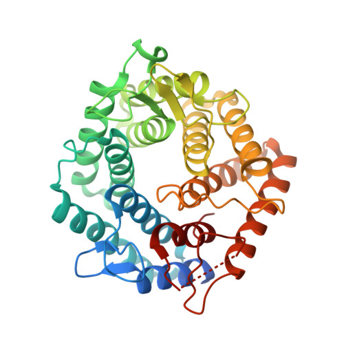
5AGD

An inactive (D125N) variant of the catalytic domain, BcGH76, of Bacillus circulans Aman6 in complex with alpha-1,6-mannopentaose


Experimental Data Snapshot

  • Method: X-RAY DIFFRACTION
  • Resolution: 1.20 Å
  • R-Value Free: 0.144 
  • R-Value Work: 0.121 
  • R-Value Observed: 0.122 

Starting Model: experimental
View more details

wwPDB Validation   3D Report Full Report


Ligand Structure Quality Assessment 


This is version 2.1 of the entry. See complete history


Literature

Evidence for a Boat Conformation at the Transition State of Gh76 Alpha-1,6-Mannanases- Key Enzymes in Bacterial and Fungal Mannoprotein Metabolism

Thompson, A.J.Speciale, G.Iglesias-Fernandez, J.Hakki, Z.Belz, T.Cartmell, A.Spears, R.J.Chandler, E.Temple, M.J.Stepper, J.Gilbert, H.J.Rovira, C.Williams, S.J.Davies, G.J.

(2015) Angew Chem Int Ed Engl 54: 5378

  • DOI: https://doi.org/10.1002/anie.201410502
  • Primary Citation of Related Structures:  
    4D4A, 4D4B, 4D4C, 4D4D, 5AGD

  • PubMed Abstract: 

    α-Mannosidases and α-mannanases have attracted attention for the insight they provide into nucleophilic substitution at the hindered anomeric center of α-mannosides, and the potential of mannosidase inhibitors as cellular probes and therapeutic agents. We report the conformational itinerary of the family GH76 α-mannanases studied through structural analysis of the Michaelis complex and synthesis and evaluation of novel aza/imino sugar inhibitors. A Michaelis complex in an (O) S2 conformation, coupled with distortion of an azasugar in an inhibitor complex to a high energy B2,5 conformation are rationalized through ab initio QM/MM metadynamics that show how the enzyme surface restricts the conformational landscape of the substrate, rendering the B2,5 conformation the most energetically stable on-enzyme. We conclude that GH76 enzymes perform catalysis using an itinerary that passes through (O) S2 and B2,5 (≠) conformations, information that should inspire the development of new antifungal agents.


  • Organizational Affiliation

    Department of Chemistry, University of York, Heslington, York, YO10 5DD (UK).


Macromolecules
Find similar proteins by:  (by identity cutoff)  |  3D Structure
Entity ID: 1
MoleculeChains Sequence LengthOrganismDetailsImage
ALPHA-1,6-MANNANASE
A, B
362Niallia circulansMutation(s): 2 
EC: 3.2.1.101
UniProt
Find proteins for Q9Z4P9 (Niallia circulans)
Explore Q9Z4P9 
Go to UniProtKB:  Q9Z4P9
Entity Groups  
Sequence Clusters30% Identity50% Identity70% Identity90% Identity95% Identity100% Identity
UniProt GroupQ9Z4P9
Sequence Annotations
Expand
  • Reference Sequence
Oligosaccharides

Help

Entity ID: 2
MoleculeChains Length2D Diagram Glycosylation3D Interactions
alpha-D-mannopyranose-(1-6)-alpha-D-mannopyranose-(1-6)-alpha-D-mannopyranose-(1-6)-alpha-D-mannopyranose-(1-6)-beta-D-mannopyranose
C, D
5N/A
Glycosylation Resources
GlyTouCan:  G20043ZO
GlyCosmos:  G20043ZO
GlyGen:  G20043ZO
Experimental Data & Validation

Experimental Data

  • Method: X-RAY DIFFRACTION
  • Resolution: 1.20 Å
  • R-Value Free: 0.144 
  • R-Value Work: 0.121 
  • R-Value Observed: 0.122 
  • Space Group: P 21 21 21
Unit Cell:
Length ( Å )Angle ( ˚ )
a = 83.52α = 90
b = 85.725β = 90
c = 102.613γ = 90
Software Package:
Software NamePurpose
REFMACrefinement
XDSdata reduction
Aimlessdata scaling
REFMACphasing

Structure Validation

View Full Validation Report


Ligand Structure Quality Assessment 


Entry History 

Revision History  (Full details and data files)

  • Version 1.0: 2015-03-25
    Type: Initial release
  • Version 1.1: 2015-04-01
    Changes: Database references
  • Version 1.2: 2015-05-06
    Changes: Database references
  • Version 1.3: 2017-07-12
    Changes: Derived calculations
  • Version 2.0: 2020-07-29
    Type: Remediation
    Reason: Carbohydrate remediation
    Changes: Atomic model, Data collection, Derived calculations, Other, Structure summary
  • Version 2.1: 2024-05-01
    Changes: Data collection, Database references, Derived calculations, Refinement description, Structure summary