6CQF

Crystal structure of HPK1 in complex an inhibitor G1858


Experimental Data Snapshot

  • Method: X-RAY DIFFRACTION
  • Resolution: 2.25 Å
  • R-Value Free: 
    0.255 (Depositor), 0.260 (DCC) 
  • R-Value Work: 
    0.208 (Depositor), 0.220 (DCC) 
  • R-Value Observed: 
    0.210 (Depositor) 

wwPDB Validation   3D Report Full Report


Ligand Structure Quality Assessment 

Created with Raphaël 2.3.0Worse 01 BetterLigand structure goodness of fit to experimental dataBest fitted F97Click on this verticalbar to view details

This is version 1.2 of the entry. See complete history


Literature

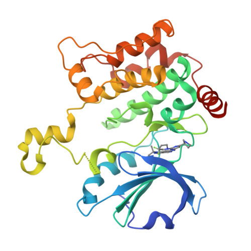
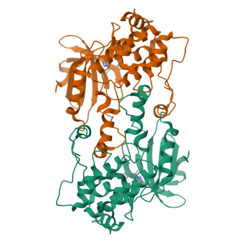
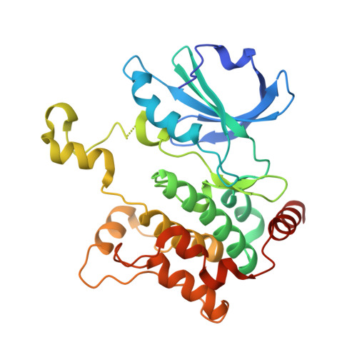
Hematopoietic Progenitor Kinase-1 Structure in a Domain-Swapped Dimer.

Wu, P.Sneeringer, C.J.Pitts, K.E.Day, E.S.Chan, B.K.Wei, B.Lehoux, I.Mortara, K.Li, H.Wu, J.Franke, Y.Moffat, J.G.Grogan, J.L.Heffron, T.P.Wang, W.

(2019) Structure 27: 125-133.e4

  • DOI: https://doi.org/10.1016/j.str.2018.10.025
  • Primary Citation of Related Structures:  
    6CQD, 6CQE, 6CQF

  • PubMed Abstract: 

    Enhancement of antigen-specific T cell immunity has shown significant therapeutic benefit in infectious diseases and cancer. Hematopoietic progenitor kinase-1 (HPK1) is a negative-feedback regulator of T cell receptor signaling, which dampens T cell proliferation and effector function. A recent report showed that a catalytic dead mutant of HPK1 phenocopies augmented T cell responses observed in HPK1-knockout mice, indicating that kinase activity is critical for function. We evaluated active and inactive mutants and determined crystal structures of HPK1 kinase domain (HPK1-KD) in apo and ligand bound forms. In all structures HPK1-KD displays a rare domain-swapped dimer, in which the activation segment comprises a well-conserved dimer interface. Biophysical measurements show formation of dimer in solution. The activation segment adopts an α-helical structure which exhibits distinct orientations in active and inactive states. This face-to-face configuration suggests that the domain-swapped dimer may possess alternative selectivity for certain substrates of HPK1 under relevant cellular context.


  • Organizational Affiliation

    Department of Structural Biology, Genentech, South San Francisco, CA 94080, USA.


Macromolecules
Find similar proteins by:  (by identity cutoff)  |  3D Structure
Entity ID: 1
MoleculeChains Sequence LengthOrganismDetailsImage
Mitogen-activated protein kinase kinase kinase kinase 1297Homo sapiensMutation(s): 0 
Gene Names: MAP4K1HPK1
EC: 2.7.11.1
UniProt & NIH Common Fund Data Resources
Find proteins for Q92918 (Homo sapiens)
Explore Q92918 
Go to UniProtKB:  Q92918
PHAROS:  Q92918
GTEx:  ENSG00000104814 
Entity Groups  
Sequence Clusters30% Identity50% Identity70% Identity90% Identity95% Identity100% Identity
UniProt GroupQ92918
Sequence Annotations
Expand
  • Reference Sequence
Small Molecules
Ligands 1 Unique
IDChains Name / Formula / InChI Key2D Diagram3D Interactions
F97
Query on F97

Download Ideal Coordinates CCD File 
B [auth A]N-{2-(3,3-difluoropyrrolidin-1-yl)-6-[(3R)-pyrrolidin-3-yl]pyrimidin-4-yl}-1-(propan-2-yl)-1H-pyrazolo[4,3-c]pyridin-6-amine
C21 H26 F2 N8
JKPAJCPZQIJPPS-CQSZACIVSA-N
Experimental Data & Validation

Experimental Data

  • Method: X-RAY DIFFRACTION
  • Resolution: 2.25 Å
  • R-Value Free:  0.255 (Depositor), 0.260 (DCC) 
  • R-Value Work:  0.208 (Depositor), 0.220 (DCC) 
  • R-Value Observed: 0.210 (Depositor) 
Space Group: C 2 2 21
Unit Cell:
Length ( Å )Angle ( ˚ )
a = 91.868α = 90
b = 98.803β = 90
c = 77.1γ = 90
Software Package:
Software NamePurpose
PHENIXrefinement
DENZOdata reduction
SCALEPACKdata scaling
PHASERphasing

Structure Validation

View Full Validation Report


Ligand Structure Quality Assessment 

Created with Raphaël 2.3.0Worse 01 BetterLigand structure goodness of fit to experimental dataBest fitted F97Click on this verticalbar to view details

Entry History 

Deposition Data

Revision History  (Full details and data files)

  • Version 1.0: 2018-12-19
    Type: Initial release
  • Version 1.1: 2019-01-16
    Changes: Data collection, Database references
  • Version 1.2: 2024-03-13
    Changes: Data collection, Database references