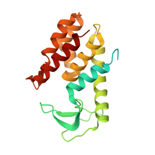
Structure of anAcinetobacterBroad-Range Prophage Endolysin Reveals a C-Terminal alpha-Helix with the Proposed Role in Activity against Live Bacterial Cells.
Sykilinda, N.N., Nikolaeva, A.Y., Shneider, M.M., Mishkin, D.V., Patutin, A.A., Popov, V.O., Boyko, K.M., Klyachko, N.L., Miroshnikov, K.A.(2018) Viruses 10
- PubMed: 29882827
- DOI: https://doi.org/10.3390/v10060309
- Primary Citation of Related Structures:
6ET6 - PubMed Abstract:
Proteins that include enzymatic domain degrading the bacterial cell wall and a domain providing transport through the bacterial outer membrane are considered as prospective compounds to combat pathogenic Gram-negative bacteria. This paper presents an isolation and study of an enzyme of this class naturally encoded in the prophage region of Acinetobacter baumannii AB 5075 genome. Recombinant protein expressed in E. coli exhibits an antimicrobial activity with respect to live cultures of Gram-negative bacteria reducing the population of viable bacteria by 1.5⁻2 log colony forming units (CFU)/mL. However the protein becomes rapidly inactivated and enables the bacteria to restore the population. AcLys structure determined by X-ray crystallography reveals a predominantly α—helical fold similar to bacteriophage P22 lysozyme. The С-terminal part of AcLys polypeptide chains forms an α—helix enriched by Lys and Arg residues exposed outside of the protein globule. Presumably this type of structure of the C-terminal α—helix has evolved evolutionally enabling the endolysin to pass the inner membrane during the host lysis or, potentially, to penetrate the outer membrane of the Gram-negative bacteria.
- Shemyakin-Ovchinnikov Institute of Bioorganic Chemistry, Russian Academy of Sciences, Moscow 117997, Russia. sykilinda@mail.ru.
Organizational Affiliation:


