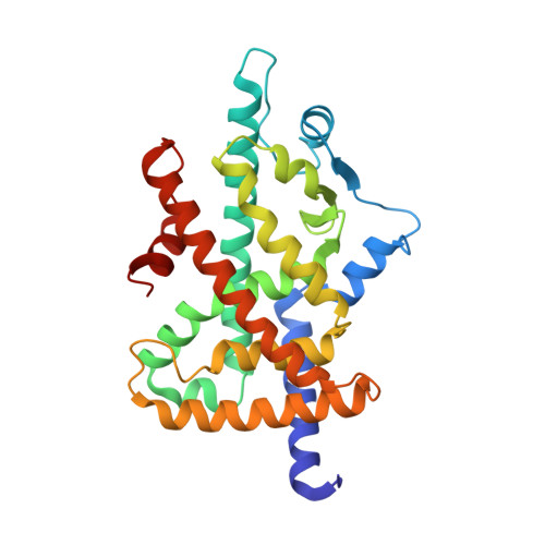
Recurrent activating mutations of PPAR gamma associated with luminal bladder tumors.
Rochel, N., Krucker, C., Coutos-Thevenot, L., Osz, J., Zhang, R., Guyon, E., Zita, W., Vanthong, S., Hernandez, O.A., Bourguet, M., Badawy, K.A., Dufour, F., Peluso-Iltis, C., Heckler-Beji, S., Dejaegere, A., Kamoun, A., de Reynies, A., Neuzillet, Y., Rebouissou, S., Beraud, C., Lang, H., Massfelder, T., Allory, Y., Cianferani, S., Stote, R.H., Radvanyi, F., Bernard-Pierrot, I.(2019) Nat Commun 10: 253-253
- PubMed: 30651555
- DOI: https://doi.org/10.1038/s41467-018-08157-y
- Primary Citation of Related Structures:
6FZF, 6FZG, 6FZJ, 6FZP, 6FZY - PubMed Abstract:
The upregulation of PPARγ/RXRα transcriptional activity has emerged as a key event in luminal bladder tumors. It renders tumor cell growth PPARγ-dependent and modulates the tumor microenvironment to favor escape from immuno-surveillance. The activation of the pathway has been linked to PPARG gains/amplifications resulting in PPARγ overexpression and to recurrent activating point mutations of RXRα. Here, we report recurrent mutations of PPARγ that also activate the PPARγ/RXRα pathway, conferring PPARγ-dependency and supporting a crucial role of PPARγ in luminal bladder cancer. These mutations are found throughout the protein-including N-terminal, DNA-binding and ligand-binding domains-and most of them enhance protein activity. Structure-function studies of PPARγ variants with mutations in the ligand-binding domain allow identifying structural elements that underpin their gain-of-function. Our study reveals genomic alterations of PPARG that lead to pro-tumorigenic PPARγ/RXRα pathway activation in luminal bladder tumors and may open the way towards alternative options for treatment.
- Institut de Génétique et de Biologie Moléculaire et Cellulaire (IGBMC), Institut National de La Santé et de La Recherche Médicale (INSERM), U1258/Centre National de Recherche Scientifique (CNRS), UMR7104/Université de Strasbourg, 67404 Illkirch, France. rochel@igbmc.fr.
Organizational Affiliation:


