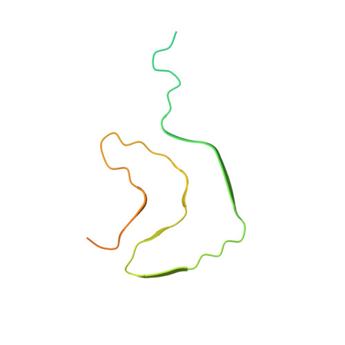
Cryo-EM structure of alpha-synuclein fibrils.
Guerrero-Ferreira, R., Taylor, N.M., Mona, D., Ringler, P., Lauer, M.E., Riek, R., Britschgi, M., Stahlberg, H.(2018) Elife 7
- PubMed: 29969391
- DOI: https://doi.org/10.7554/eLife.36402
- Primary Citation of Related Structures:
6H6B - PubMed Abstract:
Parkinson's disease is a progressive neuropathological disorder that belongs to the class of synucleinopathies, in which the protein alpha-synuclein is found at abnormally high concentrations in affected neurons. Its hallmark are intracellular inclusions called Lewy bodies and Lewy neurites. We here report the structure of cytotoxic alpha-synuclein fibrils (residues 1-121), determined by cryo-electron microscopy at a resolution of 3.4 Å. Two protofilaments form a polar fibril composed of staggered β-strands. The backbone of residues 38 to 95, including the fibril core and the non-amyloid component region, are well resolved in the EM map. Residues 50-57, containing three of the mutation sites associated with familial synucleinopathies, form the interface between the two protofilaments and contribute to fibril stability. A hydrophobic cleft at one end of the fibril may have implications for fibril elongation, and invites for the design of molecules for diagnosis and treatment of synucleinopathies.
- Center for Cellular Imaging and NanoAnalytics, Biozentrum, University of Basel, Basel, Switzerland.
Organizational Affiliation: