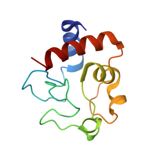
6N1O | pdb_00006n1o

Oxidized rat cytochrome c mutant (S47E)


Experimental Data Snapshot

  • Method: X-RAY DIFFRACTION
  • Resolution: 1.55 Å
  • R-Value Free: 
    0.192 (Depositor), 0.200 (DCC) 
  • R-Value Work: 
    0.166 (Depositor), 0.180 (DCC) 
  • R-Value Observed: 
    0.168 (Depositor) 

Starting Model: experimental
View more details

wwPDB Validation   3D Report Full Report


Ligand Structure Quality Assessment 

Created with Raphaël 2.3.0Worse 01 BetterLigand structure goodness of fit to experimental dataBest fitted HECClick on this verticalbar to view details

This is version 1.4 of the entry. See complete history


Literature

Serine-47 phosphorylation of cytochromecin the mammalian brain regulates cytochromecoxidase and caspase-3 activity.

Kalpage, H.A.Vaishnav, A.Liu, J.Varughese, A.Wan, J.Turner, A.A.Ji, Q.Zurek, M.P.Kapralov, A.A.Kagan, V.E.Brunzelle, J.S.Recanati, M.A.Grossman, L.I.Sanderson, T.H.Lee, I.Salomon, A.R.Edwards, B.F.P.Huttemann, M.

(2019) FASEB J 33: 13503-13514

  • DOI: https://doi.org/10.1096/fj.201901120R
  • Primary Citation of Related Structures:  
    5C0Z, 6N1O

  • PubMed Abstract: 

    Cytochrome c (Cyt c ) is a multifunctional protein that operates as an electron carrier in the mitochondrial electron transport chain and plays a key role in apoptosis. We have previously shown that tissue-specific phosphorylations of Cyt c in the heart, liver, and kidney play an important role in the regulation of cellular respiration and cell death. Here, we report that Cyt c purified from mammalian brain is phosphorylated on S47 and that this phosphorylation is lost during ischemia. We have characterized the functional effects in vitro using phosphorylated Cyt c purified from pig brain tissue and a recombinant phosphomimetic mutant (S47E). We crystallized S47E phosphomimetic Cyt c at 1.55 Å and suggest that it spatially matches S47-phosphorylated Cyt c , making it a good model system. Both S47-phosphorylated and phosphomimetic Cyt c showed a lower oxygen consumption rate in reaction with isolated Cyt c oxidase, which we propose maintains intermediate mitochondrial membrane potentials under physiologic conditions, thus minimizing production of reactive oxygen species. S47-phosphorylated and phosphomimetic Cyt c showed lower caspase-3 activity. Furthermore, phosphomimetic Cyt c had decreased cardiolipin peroxidase activity and is more stable in the presence of H 2 O 2 . Our data suggest that S47 phosphorylation of Cyt c is tissue protective and promotes cell survival in the brain.-Kalpage, H. A., Vaishnav, A., Liu, J., Varughese, A., Wan, J., Turner, A. A., Ji, Q., Zurek, M. P., Kapralov, A. A., Kagan, V. E., Brunzelle, J. S., Recanati, M.-A., Grossman, L. I., Sanderson, T. H., Lee, I., Salomon, A. R., Edwards, B. F. P, Hüttemann, M. Serine-47 phosphorylation of cytochrome c in the mammalian brain regulates cytochrome c oxidase and caspase-3 activity.


  • Organizational Affiliation

    Center for Molecular Medicine and Genetics, Wayne State University, Detroit, Michigan, USA.


Macromolecules
Find similar proteins by:  (by identity cutoff)  |  3D Structure
Entity ID: 1
MoleculeChains Sequence LengthOrganismDetailsImage
Cytochrome c, somatic
A, B, C, D
104Rattus norvegicusMutation(s): 1 
Gene Names: Cycs
UniProt
Find proteins for P62898 (Rattus norvegicus)
Explore P62898 
Go to UniProtKB:  P62898
Entity Groups  
Sequence Clusters30% Identity50% Identity70% Identity90% Identity95% Identity100% Identity
UniProt GroupP62898
Sequence Annotations
Expand
  • Reference Sequence
Small Molecules
Ligands 3 Unique
IDChains Name / Formula / InChI Key2D Diagram3D Interactions
HEC
Query on HEC

Download Ideal Coordinates CCD File 
E [auth A],
I [auth B],
L [auth C],
N [auth D]
HEME C
C34 H34 Fe N4 O4
HXQIYSLZKNYNMH-LJNAALQVSA-N
FC6
Query on FC6

Download Ideal Coordinates CCD File 
H [auth A],
J [auth B],
K [auth B],
M [auth C]
HEXACYANOFERRATE(3-)
C6 Fe N6
HCMVSLMENOCDCK-UHFFFAOYSA-N
EDO
Query on EDO

Download Ideal Coordinates CCD File 
F [auth A],
G [auth A]
1,2-ETHANEDIOL
C2 H6 O2
LYCAIKOWRPUZTN-UHFFFAOYSA-N
Experimental Data & Validation

Experimental Data

  • Method: X-RAY DIFFRACTION
  • Resolution: 1.55 Å
  • R-Value Free:  0.192 (Depositor), 0.200 (DCC) 
  • R-Value Work:  0.166 (Depositor), 0.180 (DCC) 
  • R-Value Observed: 0.168 (Depositor) 
Space Group: P 1
Unit Cell:
Length ( Å )Angle ( ˚ )
a = 34.705α = 109.89
b = 52.357β = 92.66
c = 61.819γ = 91.15
Software Package:
Software NamePurpose
XDSdata reduction
Aimlessdata scaling
PHASERphasing
REFMACrefinement
PDB_EXTRACTdata extraction

Structure Validation

View Full Validation Report


Ligand Structure Quality Assessment 

Created with Raphaël 2.3.0Worse 01 BetterLigand structure goodness of fit to experimental dataBest fitted HECClick on this verticalbar to view details

Entry History & Funding Information

Deposition Data


Funding OrganizationLocationGrant Number
National Institutes of Health/National Human Genome Research Institute (NIH/NHGRI)United StatesGM1166807

Revision History  (Full details and data files)

  • Version 1.0: 2019-10-23
    Type: Initial release
  • Version 1.1: 2019-12-11
    Changes: Database references
  • Version 1.2: 2019-12-18
    Changes: Author supporting evidence
  • Version 1.3: 2023-10-11
    Changes: Data collection, Database references, Derived calculations, Refinement description
  • Version 1.4: 2024-11-13
    Changes: Structure summary