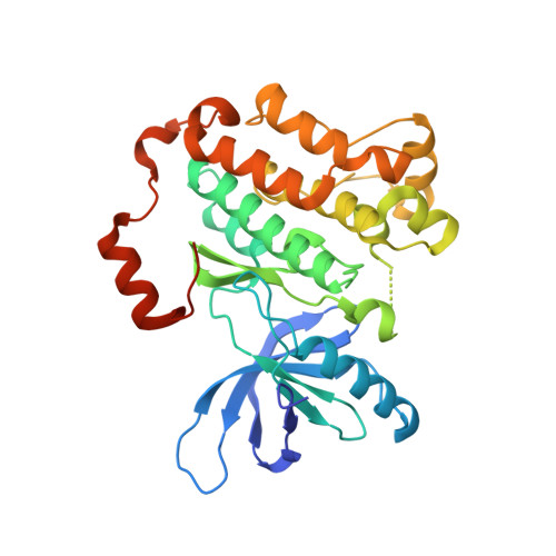
6P1D

Crystal structure of EGFR with mutant-selective dihydrodibenzodiazepinone allosteric inhibitor


Experimental Data Snapshot

  • Method: X-RAY DIFFRACTION
  • Resolution: 2.40 Å
  • R-Value Free: 0.213 
  • R-Value Work: 0.195 
  • R-Value Observed: 0.195 

Starting Model: experimental
View more details

wwPDB Validation   3D Report Full Report


Ligand Structure Quality Assessment 


This is version 1.1 of the entry. See complete history


Literature

Discovery and Optimization of Dibenzodiazepinones as Allosteric Mutant-Selective EGFR Inhibitors.

De Clercq, D.J.H.Heppner, D.E.To, C.Jang, J.Park, E.Yun, C.H.Mushajiang, M.Shin, B.H.Gero, T.W.Scott, D.A.Janne, P.A.Eck, M.J.Gray, N.S.

(2019) ACS Med Chem Lett 10: 1549-1553

  • DOI: https://doi.org/10.1021/acsmedchemlett.9b00381
  • Primary Citation of Related Structures:  
    6P1D, 6P1L, 6P8Q

  • PubMed Abstract: 

    Allosteric kinase inhibitors represent a promising new therapeutic strategy for targeting kinases harboring oncogenic driver mutations in cancers. Here, we report the discovery, optimization, and structural characterization of allosteric mutant-selective EGFR inhibitors comprising a 5,10-dihydro-11 H -dibenzo[ b , e ][1,4]diazepin-11-one scaffold. Our structure-based medicinal chemistry effort yielded an inhibitor ( 3 ) of the EGFR(L858R/T790M) and EGFR(L858R/T790M/C797S) mutants with an IC 50 of ∼10 nM and high selectivity, as assessed by kinome profiling. Further efforts to develop allosteric dibenzodiazepinone inhibitors may serve as the basis for new therapeutic options for targeting drug-resistant EGFR mutations.


  • Organizational Affiliation

    Department of Cancer Biology, Dana-Farber Cancer Institute, Boston, Massachusetts 02215, United States.


Macromolecules
Find similar proteins by:  (by identity cutoff)  |  3D Structure
Entity ID: 1
MoleculeChains Sequence LengthOrganismDetailsImage
Epidermal growth factor receptorA [auth D],
B [auth A],
C [auth B],
D [auth C]
327Homo sapiensMutation(s): 0 
Gene Names: EGFRERBBERBB1HER1
EC: 2.7.10.1
UniProt & NIH Common Fund Data Resources
Find proteins for P00533 (Homo sapiens)
Explore P00533 
Go to UniProtKB:  P00533
PHAROS:  P00533
GTEx:  ENSG00000146648 
Entity Groups  
Sequence Clusters30% Identity50% Identity70% Identity90% Identity95% Identity100% Identity
UniProt GroupP00533
Sequence Annotations
Expand
  • Reference Sequence
Small Molecules
Ligands 3 Unique
IDChains Name / Formula / InChI Key2D Diagram3D Interactions
ANP
Query on ANP

Download Ideal Coordinates CCD File 
F [auth D],
H [auth A],
J [auth B],
N [auth C]
PHOSPHOAMINOPHOSPHONIC ACID-ADENYLATE ESTER
C10 H17 N6 O12 P3
PVKSNHVPLWYQGJ-KQYNXXCUSA-N
NQ1 (Subject of Investigation/LOI)
Query on NQ1

Download Ideal Coordinates CCD File 
E [auth D],
K [auth B],
M [auth C]
10-benzyl-8-fluoro-5,10-dihydro-11H-dibenzo[b,e][1,4]diazepin-11-one
C20 H15 F N2 O
NKFCUHMOEKHJLB-UHFFFAOYSA-N
MG
Query on MG

Download Ideal Coordinates CCD File 
G [auth D],
I [auth A],
L [auth B],
O [auth C]
MAGNESIUM ION
Mg
JLVVSXFLKOJNIY-UHFFFAOYSA-N
Experimental Data & Validation

Experimental Data

  • Method: X-RAY DIFFRACTION
  • Resolution: 2.40 Å
  • R-Value Free: 0.213 
  • R-Value Work: 0.195 
  • R-Value Observed: 0.195 
  • Space Group: P 1 21 1
Unit Cell:
Length ( Å )Angle ( ˚ )
a = 71.946α = 90
b = 101.812β = 101.04
c = 86.51γ = 90
Software Package:
Software NamePurpose
PHENIXrefinement
Aimlessdata scaling
PDB_EXTRACTdata extraction
PHASERphasing
Cootmodel building
DIALSdata reduction

Structure Validation

View Full Validation Report


Ligand Structure Quality Assessment 


Entry History & Funding Information

Deposition Data


Funding OrganizationLocationGrant Number
National Institutes of Health/National Cancer Institute (NIH/NCI)United States--

Revision History  (Full details and data files)

  • Version 1.0: 2019-12-18
    Type: Initial release
  • Version 1.1: 2023-10-11
    Changes: Data collection, Database references, Derived calculations, Refinement description