6QGT

The carbon monoxide inhibition of F420-reducing [NiFe] hydrogenase complex from Methanosarcina barkeri


Experimental Data Snapshot

  • Method: X-RAY DIFFRACTION
  • Resolution: 1.99 Å
  • R-Value Free: 0.209 
  • R-Value Work: 0.169 
  • R-Value Observed: 0.170 

Starting Model: experimental
View more details

wwPDB Validation   3D Report Full Report


Ligand Structure Quality Assessment 


This is version 1.3 of the entry. See complete history


Literature

X-ray Crystallography and Vibrational Spectroscopy Reveal the Key Determinants of Biocatalytic Dihydrogen Cycling by [NiFe] Hydrogenases.

Ilina, Y.Lorent, C.Katz, S.Jeoung, J.H.Shima, S.Horch, M.Zebger, I.Dobbek, H.

(2019) Angew Chem Int Ed Engl 58: 18710-18714

  • DOI: https://doi.org/10.1002/anie.201908258
  • Primary Citation of Related Structures:  
    6QGR, 6QGT, 6QII

  • PubMed Abstract: 

    [NiFe] hydrogenases are complex model enzymes for the reversible cleavage of dihydrogen (H 2 ). However, structural determinants of efficient H 2 binding to their [NiFe] active site are not properly understood. Here, we present crystallographic and vibrational-spectroscopic insights into the unexplored structure of the H 2 -binding [NiFe] intermediate. Using an F 420 -reducing [NiFe]-hydrogenase from Methanosarcina barkeri as a model enzyme, we show that the protein backbone provides a strained chelating scaffold that tunes the [NiFe] active site for efficient H 2 binding and conversion. The protein matrix also directs H 2 diffusion to the [NiFe] site via two gas channels and allows the distribution of electrons between functional protomers through a subunit-bridging FeS cluster. Our findings emphasize the relevance of an atypical Ni coordination, thereby providing a blueprint for the design of bio-inspired H 2 -conversion catalysts.


  • Organizational Affiliation

    Institut für Biologie, Strukturbiologie/Biochemie, Humboldt-Universität zu Berlin, Philippstraße 13, 10115, Berlin, Germany.


Macromolecules
Find similar proteins by:  (by identity cutoff)  |  3D Structure
Entity ID: 1
MoleculeChains Sequence LengthOrganismDetailsImage
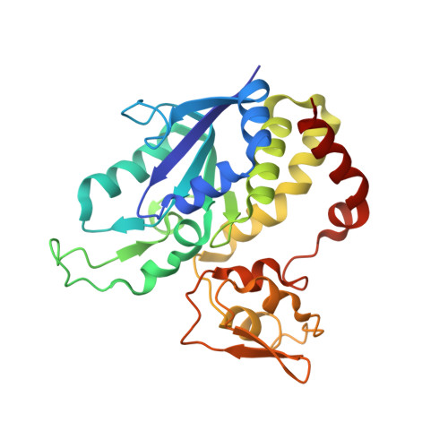
Coenzyme F420 hydrogenase subunit gammaA [auth G]253Methanosarcina barkeri MSMutation(s): 0 
EC: 1.12.98.1
UniProt
Find proteins for A0A0E3LP72 (Methanosarcina barkeri MS)
Explore A0A0E3LP72 
Go to UniProtKB:  A0A0E3LP72
Entity Groups  
Sequence Clusters30% Identity50% Identity70% Identity90% Identity95% Identity100% Identity
UniProt GroupA0A0E3LP72
Sequence Annotations
Expand
  • Reference Sequence
Find similar proteins by:  (by identity cutoff)  |  3D Structure
Entity ID: 2
MoleculeChains Sequence LengthOrganismDetailsImage
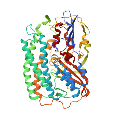
F420-reducing hydrogenase, subunit alphaB [auth A]438Methanosarcina barkeri MSMutation(s): 0 
EC: 1.12.98.1
UniProt
Find proteins for A0A0E3QYL7 (Methanosarcina barkeri MS)
Explore A0A0E3QYL7 
Go to UniProtKB:  A0A0E3QYL7
Entity Groups  
Sequence Clusters30% Identity50% Identity70% Identity90% Identity95% Identity100% Identity
UniProt GroupA0A0E3QYL7
Sequence Annotations
Expand
  • Reference Sequence
Find similar proteins by:  (by identity cutoff)  |  3D Structure
Entity ID: 3
MoleculeChains Sequence LengthOrganismDetailsImage
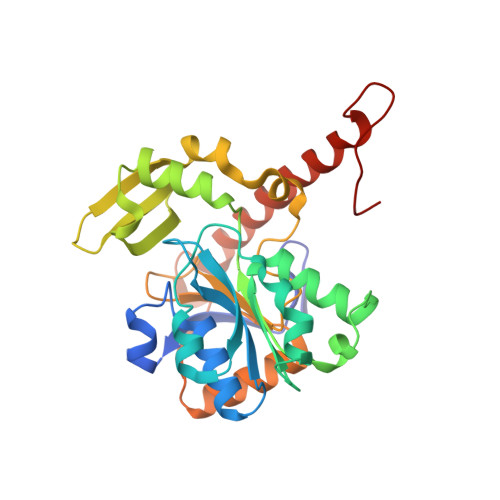
Coenzyme F420 hydrogenase subunit betaC [auth B]291Methanosarcina barkeri MSMutation(s): 0 
EC: 1.12.98.1
UniProt
Find proteins for A0A0E3QWH3 (Methanosarcina barkeri MS)
Explore A0A0E3QWH3 
Go to UniProtKB:  A0A0E3QWH3
Entity Groups  
Sequence Clusters30% Identity50% Identity70% Identity90% Identity95% Identity100% Identity
UniProt GroupA0A0E3QWH3
Sequence Annotations
Expand
  • Reference Sequence
Small Molecules
Ligands 9 Unique
IDChains Name / Formula / InChI Key2D Diagram3D Interactions
FAD
Query on FAD

Download Ideal Coordinates CCD File 
Q [auth B]FLAVIN-ADENINE DINUCLEOTIDE
C27 H33 N9 O15 P2
VWWQXMAJTJZDQX-UYBVJOGSSA-N
SF4
Query on SF4

Download Ideal Coordinates CCD File 
D [auth G],
E [auth G],
F [auth G],
R [auth B]
IRON/SULFUR CLUSTER
Fe4 S4
LJBDFODJNLIPKO-UHFFFAOYSA-N
J52
Query on J52

Download Ideal Coordinates CCD File 
K [auth A]dicyano-(oxidaniumylidynemethylnickelio)-(oxidanylidenemethylidene)iron
C4 Fe N2 Ni O2
MJFCHCXJXOCHFY-UHFFFAOYSA-N
FES
Query on FES

Download Ideal Coordinates CCD File 
G
FE2/S2 (INORGANIC) CLUSTER
Fe2 S2
NIXDOXVAJZFRNF-UHFFFAOYSA-N
144
Query on 144

Download Ideal Coordinates CCD File 
H [auth G],
N [auth A]
TRIS-HYDROXYMETHYL-METHYL-AMMONIUM
C4 H12 N O3
DRDCQJADRSJFFD-UHFFFAOYSA-N
MPD
Query on MPD

Download Ideal Coordinates CCD File 
O [auth A](4S)-2-METHYL-2,4-PENTANEDIOL
C6 H14 O2
SVTBMSDMJJWYQN-YFKPBYRVSA-N
BU3
Query on BU3

Download Ideal Coordinates CCD File 
I [auth G],
J [auth G],
P [auth B]
(R,R)-2,3-BUTANEDIOL
C4 H10 O2
OWBTYPJTUOEWEK-QWWZWVQMSA-N
FE
Query on FE

Download Ideal Coordinates CCD File 
L [auth A]FE (III) ION
Fe
VTLYFUHAOXGGBS-UHFFFAOYSA-N
MG
Query on MG

Download Ideal Coordinates CCD File 
M [auth A]MAGNESIUM ION
Mg
JLVVSXFLKOJNIY-UHFFFAOYSA-N
Experimental Data & Validation

Experimental Data

  • Method: X-RAY DIFFRACTION
  • Resolution: 1.99 Å
  • R-Value Free: 0.209 
  • R-Value Work: 0.169 
  • R-Value Observed: 0.170 
  • Space Group: F 2 3
Unit Cell:
Length ( Å )Angle ( ˚ )
a = 235.429α = 90
b = 235.429β = 90
c = 235.429γ = 90
Software Package:
Software NamePurpose
PHENIXrefinement
MxCuBEdata collection
XDSdata scaling
PHENIXphasing

Structure Validation

View Full Validation Report


Ligand Structure Quality Assessment 


Entry History & Funding Information

Deposition Data


Funding OrganizationLocationGrant Number
German Research FoundationGermanySPP 1927

Revision History  (Full details and data files)

  • Version 1.0: 2019-10-23
    Type: Initial release
  • Version 1.1: 2019-11-06
    Changes: Data collection, Database references
  • Version 1.2: 2019-12-18
    Changes: Database references
  • Version 1.3: 2024-01-24
    Changes: Data collection, Database references, Derived calculations, Refinement description