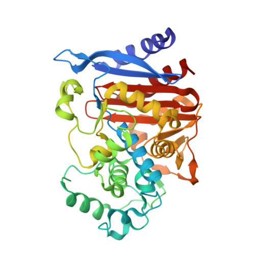
6T35

Crystal structure of AmpC from E.coli with Enmetazobactam (AAI-101)


Experimental Data Snapshot

  • Method: X-RAY DIFFRACTION
  • Resolution: 1.75 Å
  • R-Value Free: 0.184 
  • R-Value Work: 0.167 
  • R-Value Observed: 0.168 

Starting Model: experimental
View more details

wwPDB Validation   3D Report Full Report


Ligand Structure Quality Assessment 


This is version 2.1 of the entry. See complete history


Literature

Studies on enmetazobactam clarify mechanisms of widely used beta-lactamase inhibitors.

Lang, P.A.Raj, R.Tumber, A.Lohans, C.T.Rabe, P.Robinson, C.V.Brem, J.Schofield, C.J.

(2022) Proc Natl Acad Sci U S A 119: e2117310119-e2117310119

  • DOI: https://doi.org/10.1073/pnas.2117310119
  • Primary Citation of Related Structures:  
    6T35, 7B3R, 7B3S, 7B3U

  • PubMed Abstract: 

    β-Lactams are the most important class of antibacterials, but their use is increasingly compromised by resistance, most importantly via serine β-lactamase (SBL)-catalyzed hydrolysis. The scope of β-lactam antibacterial activity can be substantially extended by coadministration with a penicillin-derived SBL inhibitor (SBLi), i.e., the penam sulfones tazobactam and sulbactam, which are mechanism-based inhibitors working by acylation of the nucleophilic serine. The new SBLi enmetazobactam, an N-methylated tazobactam derivative, has recently completed clinical trials. Biophysical studies on the mechanism of SBL inhibition by enmetazobactam reveal that it inhibits representatives of all SBL classes without undergoing substantial scaffold fragmentation, a finding that contrasts with previous reports on SBL inhibition by tazobactam and sulbactam. We therefore reinvestigated the mechanisms of tazobactam and sulbactam using mass spectrometry under denaturing and nondenaturing conditions, X-ray crystallography, and NMR spectroscopy. The results imply that the reported extensive fragmentation of penam sulfone–derived acyl–enzyme complexes does not substantially contribute to SBL inhibition. In addition to observation of previously identified inhibitor-induced SBL modifications, the results reveal that prolonged reaction of penam sulfones with SBLs can induce dehydration of the nucleophilic serine to give a dehydroalanine residue that undergoes reaction to give a previously unobserved lysinoalanine cross-link. The results clarify the mechanisms of action of widely clinically used SBLi, reveal limitations on the interpretation of mass spectrometry studies concerning mechanisms of SBLi, and will inform the development of new SBLi working by reaction to form hydrolytically stable acyl–enzyme complexes.


  • Organizational Affiliation

    Chemistry Research Laboratory, Department of Chemistry, University of Oxford, Oxford OX1 3TA, United Kingdom.


Macromolecules
Find similar proteins by:  (by identity cutoff)  |  3D Structure
Entity ID: 1
MoleculeChains Sequence LengthOrganismDetailsImage
Beta-lactamase358Escherichia coli K-12Mutation(s): 0 
Gene Names: ampCampAb4150JW4111
EC: 3.5.2.6
UniProt
Find proteins for P00811 (Escherichia coli (strain K12))
Explore P00811 
Go to UniProtKB:  P00811
Entity Groups  
Sequence Clusters30% Identity50% Identity70% Identity90% Identity95% Identity100% Identity
UniProt GroupP00811
Sequence Annotations
Expand
  • Reference Sequence
Small Molecules
Ligands 5 Unique
IDChains Name / Formula / InChI Key2D Diagram3D Interactions
M9W (Subject of Investigation/LOI)
Query on M9W

Download Ideal Coordinates CCD File 
E [auth A]Enmetazobactam derived trans-enamine adduct
C11 H17 N4 O6 S
VVXNTAXAJHAELU-UEVFTQGLSA-O
PEG
Query on PEG

Download Ideal Coordinates CCD File 
C [auth A],
D [auth A]
DI(HYDROXYETHYL)ETHER
C4 H10 O3
MTHSVFCYNBDYFN-UHFFFAOYSA-N
SO4
Query on SO4

Download Ideal Coordinates CCD File 
B [auth A]SULFATE ION
O4 S
QAOWNCQODCNURD-UHFFFAOYSA-L
ZN
Query on ZN

Download Ideal Coordinates CCD File 
F [auth A],
G [auth A],
H [auth A]
ZINC ION
Zn
PTFCDOFLOPIGGS-UHFFFAOYSA-N
CL
Query on CL

Download Ideal Coordinates CCD File 
I [auth A]CHLORIDE ION
Cl
VEXZGXHMUGYJMC-UHFFFAOYSA-M
Experimental Data & Validation

Experimental Data

  • Method: X-RAY DIFFRACTION
  • Resolution: 1.75 Å
  • R-Value Free: 0.184 
  • R-Value Work: 0.167 
  • R-Value Observed: 0.168 
  • Space Group: P 43 3 2
Unit Cell:
Length ( Å )Angle ( ˚ )
a = 138α = 90
b = 138β = 90
c = 138γ = 90
Software Package:
Software NamePurpose
XDSdata reduction
Aimlessdata scaling
PHASERphasing
PHENIXrefinement
PDB_EXTRACTdata extraction

Structure Validation

View Full Validation Report


Ligand Structure Quality Assessment 


Entry History & Funding Information

Deposition Data


Funding OrganizationLocationGrant Number
Medical Research Council (United Kingdom)United KingdomMRF-145-0004-TPG-AVISO

Revision History  (Full details and data files)

  • Version 1.0: 2020-11-18
    Type: Initial release
  • Version 1.1: 2022-05-11
    Changes: Database references
  • Version 1.2: 2024-01-24
    Changes: Data collection, Refinement description
  • Version 2.0: 2024-03-27
    Changes: Data collection, Derived calculations, Non-polymer description, Structure summary
  • Version 2.1: 2024-11-13
    Changes: Structure summary