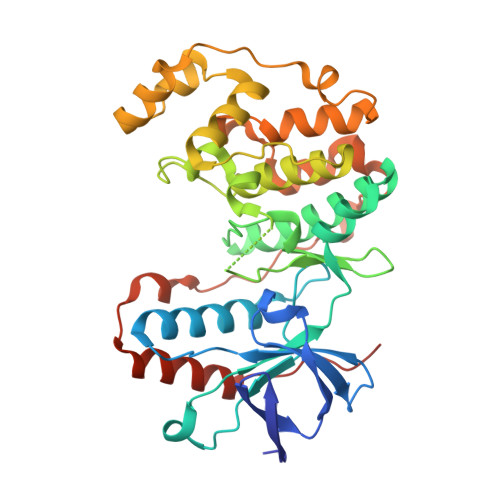
RCSB PDB - 6Y4U: Crystal structure of p38 in complex with SR65

 6Y4U

Crystal structure of p38 in complex with SR65


Experimental Data Snapshot

  • Method: X-RAY DIFFRACTION
  • Resolution: 1.86 Å
  • R-Value Free: 0.241 
  • R-Value Work: 0.190 
  • R-Value Observed: 0.192 

Starting Model: experimental
View more details

wwPDB Validation   3D Report Full Report


Ligand Structure Quality Assessment 

Created with Raphaël 2.3.0Worse 01 BetterLigand structure goodness of fit to experimental dataBest fitted O98Click on this verticalbar to view details

This is version 1.2 of the entry. See complete history


Literature

Selective targeting of the alpha C and DFG-out pocket in p38 MAPK.

Rohm, S.Schroder, M.Dwyer, J.E.Widdowson, C.S.Chaikuad, A.Berger, B.T.Joerger, A.C.Kramer, A.Harbig, J.Dauch, D.Kudolo, M.Laufer, S.Bagley, M.C.Knapp, S.

(2020) Eur J Med Chem 208: 112721-112721

  • DOI: https://doi.org/10.1016/j.ejmech.2020.112721
  • Primary Citation of Related Structures:  
    6Y4T, 6Y4U, 6Y4V, 6Y4W, 6Y4X, 6Y6V, 6YJC, 6YK7, 6ZWP, 6ZWR

  • PubMed Abstract: 

    The p38 MAPK cascade is a key signaling pathway linked to a multitude of physiological functions and of central importance in inflammatory and autoimmune diseases. Although studied extensively, little is known about how conformation-specific inhibitors alter signaling outcomes. Here, we have explored the highly dynamic back pocket of p38 MAPK with allosteric urea fragments. However, screening against known off-targets showed that these fragments maintained the selectivity issues of their parent compound BIRB-796, while combination with the hinge-binding motif of VPC-00628 greatly enhanced inhibitor selectivity. Further efforts focused therefore on the exploration of the αC-out pocket of p38 MAPK, yielding compound 137 as a highly selective type-II inhibitor. Even though 137 is structurally related to a recent p38 type-II chemical probe, SR-318, the data presented here provide valuable insights into back-pocket interactions that are not addressed in SR-318 and it provides an alternative chemical tool with good cellular activity targeting also the p38 back pocket.


  • Organizational Affiliation

    Johann Wolfgang Goethe University, Institute of Pharmaceutical Chemistry, Max-von-Laue-Str. 9, 60438, Frankfurt Am Main, Germany; Buchmann Institute for Molecular Life Sciences and Structural Genomics Consortium (SGC), Max-von-Laue-Str. 15, 60438, Frankfurt Am Main, Germany.


Macromolecules
Find similar proteins by:  (by identity cutoff)  |  3D Structure
Entity ID: 1
MoleculeChains Sequence LengthOrganismDetailsImage
Mitogen-activated protein kinase 14361Mus musculusMutation(s): 0 
Gene Names: Mapk14Crk1Csbp1Csbp2
EC: 2.7.11.24
UniProt
Find proteins for P47811 (Mus musculus)
Explore P47811 
Go to UniProtKB:  P47811
Entity Groups  
Sequence Clusters30% Identity50% Identity70% Identity90% Identity95% Identity100% Identity
UniProt GroupP47811
Sequence Annotations
Expand
  • Reference Sequence
Experimental Data & Validation

Experimental Data

  • Method: X-RAY DIFFRACTION
  • Resolution: 1.86 Å
  • R-Value Free: 0.241 
  • R-Value Work: 0.190 
  • R-Value Observed: 0.192 
  • Space Group: P 21 21 21
Unit Cell:
Length ( Å )Angle ( ˚ )
a = 66.019α = 90
b = 74.919β = 90
c = 77.799γ = 90
Software Package:
Software NamePurpose
SCALAdata scaling
REFMACrefinement
PDB_EXTRACTdata extraction
iMOSFLMdata reduction
PHASERphasing

Structure Validation

View Full Validation Report


Ligand Structure Quality Assessment 

Created with Raphaël 2.3.0Worse 01 BetterLigand structure goodness of fit to experimental dataBest fitted O98Click on this verticalbar to view details

Entry History 

Deposition Data

Revision History  (Full details and data files)

  • Version 1.0: 2020-03-04
    Type: Initial release
  • Version 1.1: 2021-01-13
    Changes: Database references
  • Version 1.2: 2024-01-24
    Changes: Data collection, Database references, Refinement description