6YMI

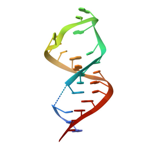
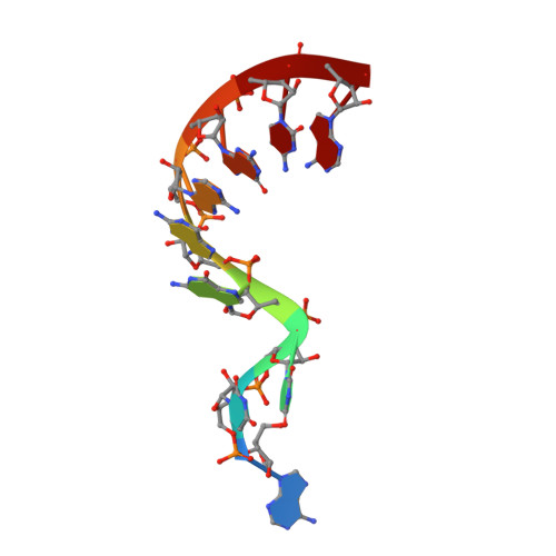
Crystal structure of the SAM-SAH riboswitch with AMP.

  • Classification: RNA
  • Organism(s): Roseobacter sp.
  • Mutation(s): No 

  • Deposited: 2020-04-08 Released: 2020-07-22 
  • Deposition Author(s): Huang, L., Lilley, D.M.J.
  • Funding Organization(s): Cancer Research UK, Ministry of Education (MoE, China)

Experimental Data Snapshot

  • Method: X-RAY DIFFRACTION
  • Resolution: 2.50 Å
  • R-Value Free: 0.242 
  • R-Value Work: 0.214 
  • R-Value Observed: 0.215 

Starting Model: experimental
View more details

wwPDB Validation   3D Report Full Report


Ligand Structure Quality Assessment 


This is version 2.2 of the entry. See complete history


Literature

Crystal structure and ligand-induced folding of the SAM/SAH riboswitch.

Huang, L.Liao, T.W.Wang, J.Ha, T.Lilley, D.M.J.

(2020) Nucleic Acids Res 48: 7545-7556

  • DOI: https://doi.org/10.1093/nar/gkaa493
  • Primary Citation of Related Structures:  
    6YL5, 6YLB, 6YMI, 6YMJ, 6YMK, 6YML, 6YMM

  • PubMed Abstract: 

    While most SAM riboswitches strongly discriminate between SAM and SAH, the SAM/SAH riboswitch responds to both ligands with similar apparent affinities. We have determined crystal structures of the SAM/SAH riboswitch bound to SAH, SAM and other variant ligands at high resolution. The riboswitch forms an H-type pseudoknot structure with coaxial alignment of the stem-loop helix (P1) and the pseudoknot helix (PK). An additional three base pairs form at the non-open end of P1, and the ligand is bound at the interface between the P1 extension and the PK helix. The adenine nucleobase is stacked into the helix and forms a trans Hoogsteen-Watson-Crick base pair with a uridine, thus becoming an integral part of the helical structure. The majority of the specific interactions are formed with the adenosine. The methionine or homocysteine chain lies in the groove making a single hydrogen bond, and there is no discrimination between the sulfonium of SAM or the thioether of SAH. Single-molecule FRET analysis reveals that the riboswitch exists in two distinct conformations, and that addition of SAM or SAH shifts the population into a stable state that likely corresponds to the form observed in the crystal. A model for translational regulation is presented whereby in the absence of ligand the riboswitch is largely unfolded, lacking the PK helix so that translation can be initiated at the ribosome binding site. But the presence of ligand stabilizes the folded conformation that includes the PK helix, so occluding the ribosome binding site and thus preventing the initiation of translation.


  • Organizational Affiliation

    Guangdong Provincial Key Laboratory of Malignant Tumor Epigenetics and Gene Regulation, Medical Research Center, Sun Yat-Sen Memorial Hospital, Sun Yat-Sen University, Guangzhou 510120, P. R. China.


Macromolecules
Find similar nucleic acids by:  (by identity cutoff)  |  3D Structure
Entity ID: 1
MoleculeChains LengthOrganismImage
Chains: A,C,F,I,M,O26Roseobacter sp.
Sequence Annotations
Expand
  • Reference Sequence

Find similar nucleic acids by:  Sequence   |   3D Structure  

Entity ID: 2
MoleculeChains LengthOrganismImage
Chains: B,D,G,J,N,P9Roseobacter sp.
Sequence Annotations
Expand
  • Reference Sequence
Small Molecules
Ligands 2 Unique
IDChains Name / Formula / InChI Key2D Diagram3D Interactions
CBV
Query on CBV

Download Ideal Coordinates CCD File 
M [auth A],
P [auth C],
R [auth F],
U [auth I],
W [auth M]
5-BROMOCYTIDINE 5'-(DIHYDROGEN PHOSPHATE)
C9 H13 Br N3 O8 P
KOHPTSGPCYTYFB-UAKXSSHOSA-N
AMP (Subject of Investigation/LOI)
Query on AMP

Download Ideal Coordinates CCD File 
N [auth A]
O [auth A]
Q [auth C]
S [auth F]
T [auth G]
N [auth A],
O [auth A],
Q [auth C],
S [auth F],
T [auth G],
V [auth I],
X [auth M],
Y [auth N],
Z [auth O]
ADENOSINE MONOPHOSPHATE
C10 H14 N5 O7 P
UDMBCSSLTHHNCD-KQYNXXCUSA-N
Experimental Data & Validation

Experimental Data

  • Method: X-RAY DIFFRACTION
  • Resolution: 2.50 Å
  • R-Value Free: 0.242 
  • R-Value Work: 0.214 
  • R-Value Observed: 0.215 
  • Space Group: C 1 2 1
Unit Cell:
Length ( Å )Angle ( ˚ )
a = 86.506α = 90
b = 147.359β = 91.364
c = 74.844γ = 90
Software Package:
Software NamePurpose
PHENIXrefinement
PHENIXrefinement
xia2data reduction
xia2data scaling
PHASERphasing

Structure Validation

View Full Validation Report


Ligand Structure Quality Assessment 


Entry History & Funding Information

Deposition Data


Funding OrganizationLocationGrant Number
Cancer Research UKUnited KingdomA18604
Ministry of Education (MoE, China)China--

Revision History  (Full details and data files)

  • Version 1.0: 2020-07-22
    Type: Initial release
  • Version 1.1: 2020-07-29
    Changes: Database references
  • Version 2.0: 2022-04-20
    Changes: Advisory, Atomic model, Data collection, Database references, Derived calculations, Structure summary
  • Version 2.1: 2023-03-08
    Changes: Structure summary
  • Version 2.2: 2024-02-07
    Changes: Data collection, Refinement description