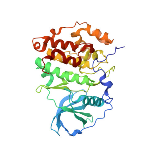
6YPG

Crystal Structure of CK2alpha with Compound 2 bound to second crystal form


Experimental Data Snapshot

  • Method: X-RAY DIFFRACTION
  • Resolution: 1.51 Å
  • R-Value Free: 0.243 
  • R-Value Work: 0.237 
  • R-Value Observed: 0.238 

Starting Model: experimental
View more details

wwPDB Validation   3D Report Full Report


Ligand Structure Quality Assessment 


This is version 1.3 of the entry. See complete history


Literature

Proposed Allosteric Inhibitors Bind to the ATP Site of CK2 alpha.

Brear, P.Ball, D.Stott, K.D'Arcy, S.Hyvonen, M.

(2020) J Med Chem 63: 12786-12798

  • DOI: https://doi.org/10.1021/acs.jmedchem.0c01173
  • Primary Citation of Related Structures:  
    6YPG, 6YPH, 6YPJ, 6YPK, 6YPN

  • PubMed Abstract: 

    CK2α is a ubiquitous, well-studied kinase that is a target for small-molecule inhibition, for treatment of cancers. While many different classes of adenosine 5'-triphosphate (ATP)-competitive inhibitors have been described for CK2α, they tend to suffer from significant off-target activity and new approaches are needed. A series of inhibitors of CK2α has recently been described as allosteric, acting at a previously unidentified binding site. Given the similarity of these inhibitors to known ATP-competitive inhibitors, we have investigated them further. In our thorough structural and biophysical analyses, we have found no evidence that these inhibitors bind to the proposed allosteric site. Rather, we report crystal structures, competitive isothermal titration calorimetry (ITC) and NMR, hydrogen-deuterium exchange (HDX) mass spectrometry, and chemoinformatic analyses that all point to these compounds binding in the ATP pocket. Comparisons of our results and experimental approach with the data presented in the original report suggest that the primary reason for the disparity is nonspecific inhibition by aggregation.


  • Organizational Affiliation

    Department of Biochemistry, University of Cambridge, Cambridge CB2 1GA, U.K.


Macromolecules
Find similar proteins by:  (by identity cutoff)  |  3D Structure
Entity ID: 1
MoleculeChains Sequence LengthOrganismDetailsImage
Casein kinase II subunit alpha328Homo sapiensMutation(s): 4 
Gene Names: CSNK2A1CK2A1
EC: 2.7.11.1
UniProt & NIH Common Fund Data Resources
Find proteins for P68400 (Homo sapiens)
Explore P68400 
Go to UniProtKB:  P68400
PHAROS:  P68400
GTEx:  ENSG00000101266 
Entity Groups  
Sequence Clusters30% Identity50% Identity70% Identity90% Identity95% Identity100% Identity
UniProt GroupP68400
Sequence Annotations
Expand
  • Reference Sequence
Small Molecules
Ligands 2 Unique
IDChains Name / Formula / InChI Key2D Diagram3D Interactions
N5Q (Subject of Investigation/LOI)
Query on N5Q

Download Ideal Coordinates CCD File 
B [auth A]4-[(4-naphthalen-2-yl-1,3-thiazol-2-yl)amino]-2-oxidanyl-benzoic acid
C20 H14 N2 O3 S
HUECFCSRZKSRJF-UHFFFAOYSA-N
ACT
Query on ACT

Download Ideal Coordinates CCD File 
C [auth A]ACETATE ION
C2 H3 O2
QTBSBXVTEAMEQO-UHFFFAOYSA-M
Experimental Data & Validation

Experimental Data

  • Method: X-RAY DIFFRACTION
  • Resolution: 1.51 Å
  • R-Value Free: 0.243 
  • R-Value Work: 0.237 
  • R-Value Observed: 0.238 
  • Space Group: P 1 21 1
Unit Cell:
Length ( Å )Angle ( ˚ )
a = 57.653α = 90
b = 45.661β = 111.05
c = 63.817γ = 90
Software Package:
Software NamePurpose
REFMACrefinement
XDSdata reduction
Aimlessdata scaling
PHASERphasing
PDB_EXTRACTdata extraction

Structure Validation

View Full Validation Report


Ligand Structure Quality Assessment 


Entry History 

Deposition Data

Revision History  (Full details and data files)

  • Version 1.0: 2020-07-15
    Type: Initial release
  • Version 1.1: 2020-11-11
    Changes: Database references
  • Version 1.2: 2020-11-25
    Changes: Database references
  • Version 1.3: 2024-01-24
    Changes: Data collection, Database references, Refinement description