6Z6F

HDAC-PC


Experimental Data Snapshot

  • Method: ELECTRON MICROSCOPY
  • Resolution: 3.11 Å
  • Aggregation State: PARTICLE 
  • Reconstruction Method: SINGLE PARTICLE 

wwPDB Validation   3D Report Full Report


This is version 1.0 of the entry. See complete history


Literature

Structural basis for the regulation of nucleosome recognition and HDAC activity by histone deacetylase assemblies.

Lee, J.H.Bollschweiler, D.Schafer, T.Huber, R.

(2021) Sci Adv 7

  • DOI: https://doi.org/10.1126/sciadv.abd4413
  • Primary Citation of Related Structures:  
    6Z6F, 6Z6H, 6Z6O, 6Z6P

  • PubMed Abstract: 

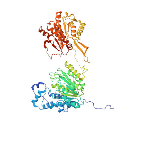
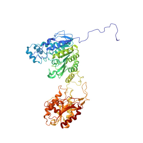
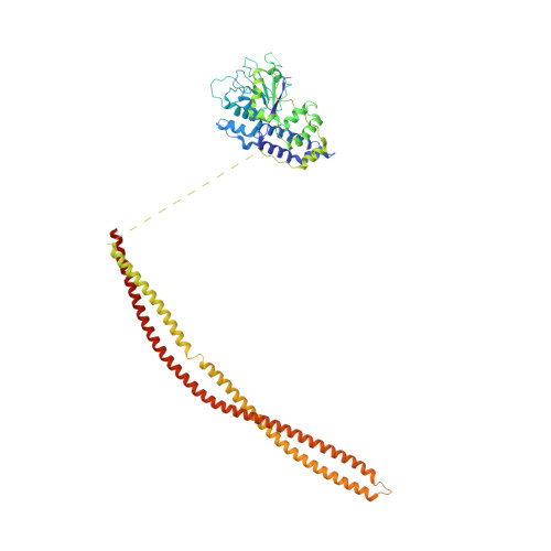
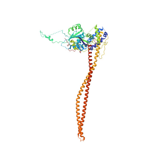
    The chromatin-modifying histone deacetylases (HDACs) remove acetyl groups from acetyl-lysine residues in histone amino-terminal tails, thereby mediating transcriptional repression. Structural makeup and mechanisms by which multisubunit HDAC complexes recognize nucleosomes remain elusive. Our cryo-electron microscopy structures of the yeast class II HDAC ensembles show that the HDAC protomer comprises a triangle-shaped assembly of stoichiometry Hda1 2 -Hda2-Hda3, in which the active sites of the Hda1 dimer are freely accessible. We also observe a tetramer of protomers, where the nucleosome binding modules are inaccessible. Structural analysis of the nucleosome-bound complexes indicates how positioning of Hda1 adjacent to histone H2B affords HDAC catalysis. Moreover, it reveals how an intricate network of multiple contacts between a dimer of protomers and the nucleosome creates a platform for expansion of the HDAC activities. Our study provides comprehensive insight into the structural plasticity of the HDAC complex and its functional mechanism of chromatin modification.


  • Organizational Affiliation

    Max Planck Institute of Biochemistry, Am Klopferspitz 18, 82152 Martinsried, Germany. hlee@biochem.mpg.de huber@biochem.mpg.de.


Macromolecules
Find similar proteins by:  (by identity cutoff)  |  3D Structure
Entity ID: 1
MoleculeChains Sequence LengthOrganismDetailsImage
Histone deacetylase HDA1661Saccharomyces cerevisiae S288CMutation(s): 0 
Gene Names: HDA1YNL021WN2819
EC: 3.5.1.98
UniProt
Find proteins for P53973 (Saccharomyces cerevisiae (strain ATCC 204508 / S288c))
Explore P53973 
Go to UniProtKB:  P53973
Entity Groups  
Sequence Clusters30% Identity50% Identity70% Identity90% Identity95% Identity100% Identity
UniProt GroupP53973
Sequence Annotations
Expand
  • Reference Sequence
Find similar proteins by:  (by identity cutoff)  |  3D Structure
Entity ID: 2
MoleculeChains Sequence LengthOrganismDetailsImage
Histone deacetylase HDA1672Saccharomyces cerevisiae S288CMutation(s): 0 
Gene Names: HDA1YNL021WN2819
EC: 3.5.1.98
UniProt
Find proteins for P53973 (Saccharomyces cerevisiae (strain ATCC 204508 / S288c))
Explore P53973 
Go to UniProtKB:  P53973
Entity Groups  
Sequence Clusters30% Identity50% Identity70% Identity90% Identity95% Identity100% Identity
UniProt GroupP53973
Sequence Annotations
Expand
  • Reference Sequence
Find similar proteins by:  (by identity cutoff)  |  3D Structure
Entity ID: 3
MoleculeChains Sequence LengthOrganismDetailsImage
HDA1 complex subunit 3,HDA1 complex subunit 3C [auth D]543Saccharomyces cerevisiae S288CMutation(s): 0 
Gene Names: HDA3PLO1YPR179C
UniProt
Find proteins for Q06623 (Saccharomyces cerevisiae (strain ATCC 204508 / S288c))
Explore Q06623 
Go to UniProtKB:  Q06623
Entity Groups  
Sequence Clusters30% Identity50% Identity70% Identity90% Identity95% Identity100% Identity
UniProt GroupQ06623
Sequence Annotations
Expand
  • Reference Sequence
Find similar proteins by:  (by identity cutoff)  |  3D Structure
Entity ID: 4
MoleculeChains Sequence LengthOrganismDetailsImage
HDA1 complex subunit 2D [auth C]629Saccharomyces cerevisiae S288CMutation(s): 0 
Gene Names: HDA2PLO2YDR295C
UniProt
Find proteins for Q06629 (Saccharomyces cerevisiae (strain ATCC 204508 / S288c))
Explore Q06629 
Go to UniProtKB:  Q06629
Entity Groups  
Sequence Clusters30% Identity50% Identity70% Identity90% Identity95% Identity100% Identity
UniProt GroupQ06629
Sequence Annotations
Expand
  • Reference Sequence
Experimental Data & Validation

Experimental Data

  • Method: ELECTRON MICROSCOPY
  • Resolution: 3.11 Å
  • Aggregation State: PARTICLE 
  • Reconstruction Method: SINGLE PARTICLE 
EM Software:
TaskSoftware PackageVersion
RECONSTRUCTIONcryoSPARC2
MODEL REFINEMENTPHENIX

Structure Validation

View Full Validation Report


Entry History & Funding Information

Deposition Data


Funding OrganizationLocationGrant Number
Max Planck SocietyGermany--

Revision History  (Full details and data files)

  • Version 1.0: 2021-02-17
    Type: Initial release