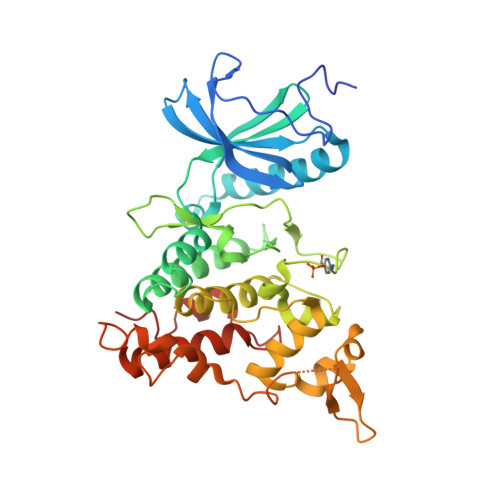
7FHT

Crystal structure of DYRK1A in complex with RD0448

  • Classification: TRANSFERASE
  • Organism(s): Homo sapiens
  • Expression System: Escherichia coli
  • Mutation(s): No 

  • Deposited: 2021-07-30 Released: 2022-03-23 
  • Deposition Author(s): Kikuchi, M., Sumida, Y., Hosoya, T., Kii, I., Umehara, T.
  • Funding Organization(s): Ministry of Education, Culture, Sports, Science and Technology (Japan), Kobayashi Foundation, Life Science Foundation of JAPAN, Tokyo Biochemical Research Foundation, Mochida Memorial Foundation for Medical and Pharmaceutical Research

Experimental Data Snapshot

  • Method: X-RAY DIFFRACTION
  • Resolution: 2.68 Å
  • R-Value Free: 0.249 
  • R-Value Work: 0.188 
  • R-Value Observed: 0.191 

Starting Model: experimental
View more details

wwPDB Validation   3D Report Full Report


Ligand Structure Quality Assessment 


This is version 1.2 of the entry. See complete history


Literature

Structure-activity relationship for the folding intermediate-selective inhibition of DYRK1A.

Miyazaki, Y.Kikuchi, M.Umezawa, K.Descamps, A.Nakamura, D.Furuie, G.Sumida, T.Saito, K.Kimura, N.Niwa, T.Sumida, Y.Umehara, T.Hosoya, T.Kii, I.

(2022) Eur J Med Chem 227: 113948-113948

  • DOI: https://doi.org/10.1016/j.ejmech.2021.113948
  • Primary Citation of Related Structures:  
    7FHS, 7FHT

  • PubMed Abstract: 

    DYRK1A phosphorylates proteins involved in neurological disorders in an intermolecular manner. Meanwhile, during the protein folding process of DYRK1A, a transitional folding intermediate catalyzes the intramolecular autophosphorylation required for the "one-off" inceptive activation and stabilization. In our previous study, a small molecule termed FINDY (1) was identified, which inhibits the folding intermediate-catalyzed intramolecular autophosphorylation of DYRK1A but not the folded state-catalyzed intermolecular phosphorylation. However, the structural features of FINDY (1) responsible for this intermediate-selective inhibition remain elusive. In this study, structural derivatives of FINDY (1) were designed and synthesized according to its predicted binding mode in the ATP pocket of DYRK1A. Quantitative structure-activity relationship (QSAR) of the derivatives revealed that the selectivity against the folding intermediate is determined by steric hindrance between the bulky hydrophobic moiety of the derivatives and the entrance to the pocket. In addition, a potent derivative 3 was identified, which inhibited the folding intermediate more strongly than FINDY (1); it was designated as dp-FINDY. Although dp-FINDY (3) did not inhibit the folded state, as well as FINDY (1), it inhibited the intramolecular autophosphorylation of DYRK1A in an in vitro cell-free protein synthesis assay. Furthermore, dp-FINDY (3) destabilized endogenous DYRK1A in HEK293 cells. This study provides structural insights into the folding intermediate-selective inhibition of DYRK1A and expands the chemical options for the design of a kinase inhibitor.


  • Organizational Affiliation

    Laboratory for Drug Target Research, Department of Agriculture, Graduate School of Science and Technology, Shinshu University, 8304 Minami-Minowa, Kami-Ina, Nagano, 399-4598, Japan.


Macromolecules
Find similar proteins by:  (by identity cutoff)  |  3D Structure
Entity ID: 1
MoleculeChains Sequence LengthOrganismDetailsImage
Dual specificity tyrosine-phosphorylation-regulated kinase 1A
A, B, C, D
361Homo sapiensMutation(s): 0 
Gene Names: DYRK1ADYRKMNBMNBH
EC: 2.7.12.1 (PDB Primary Data), 2.7.11.23 (UniProt)
UniProt & NIH Common Fund Data Resources
Find proteins for Q13627 (Homo sapiens)
Explore Q13627 
Go to UniProtKB:  Q13627
PHAROS:  Q13627
GTEx:  ENSG00000157540 
Entity Groups  
Sequence Clusters30% Identity50% Identity70% Identity90% Identity95% Identity100% Identity
UniProt GroupQ13627
Sequence Annotations
Expand
  • Reference Sequence
Experimental Data & Validation

Experimental Data

  • Method: X-RAY DIFFRACTION
  • Resolution: 2.68 Å
  • R-Value Free: 0.249 
  • R-Value Work: 0.188 
  • R-Value Observed: 0.191 
  • Space Group: P 21 21 21
Unit Cell:
Length ( Å )Angle ( ˚ )
a = 87.088α = 90
b = 87.577β = 90
c = 227.146γ = 90
Software Package:
Software NamePurpose
REFMACrefinement
XDSdata reduction
Aimlessdata scaling
MOLREPphasing

Structure Validation

View Full Validation Report


Ligand Structure Quality Assessment 


Entry History & Funding Information

Deposition Data


Funding OrganizationLocationGrant Number
Ministry of Education, Culture, Sports, Science and Technology (Japan)Japan19H02856
Ministry of Education, Culture, Sports, Science and Technology (Japan)Japan18H04568
Ministry of Education, Culture, Sports, Science and Technology (Japan)Japan17H05678
Ministry of Education, Culture, Sports, Science and Technology (Japan)Japan16H05926
Kobayashi FoundationJapan--
Life Science Foundation of JAPANJapan--
Tokyo Biochemical Research FoundationJapan--
Mochida Memorial Foundation for Medical and Pharmaceutical ResearchJapan--

Revision History  (Full details and data files)

  • Version 1.0: 2022-03-23
    Type: Initial release
  • Version 1.1: 2023-11-29
    Changes: Data collection, Refinement description
  • Version 1.2: 2024-11-13
    Changes: Structure summary