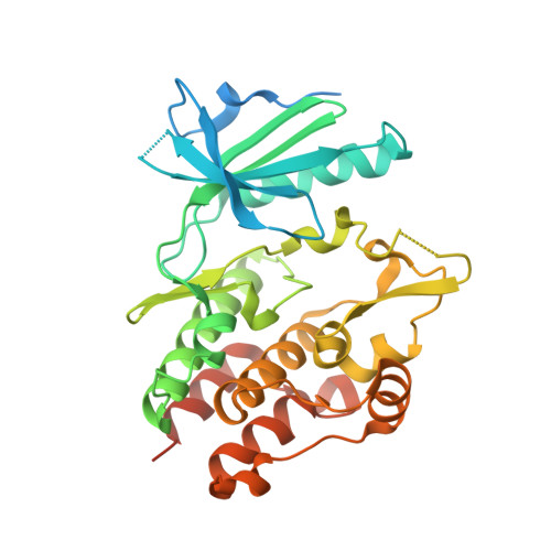
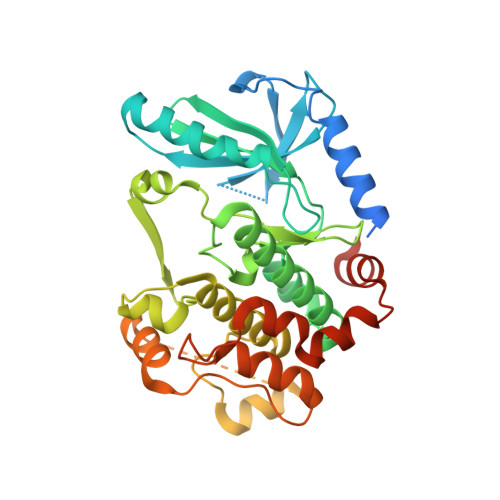
7JUU

Crystal Structure of KSR2:MEK1 in complex with AMP-PNP, and allosteric MEK inhibitor PD0325901


Experimental Data Snapshot

  • Method: X-RAY DIFFRACTION
  • Resolution: 3.19 Å
  • R-Value Free: 0.258 
  • R-Value Work: 0.241 
  • R-Value Observed: 0.242 

Starting Model: experimental
View more details

wwPDB Validation   3D Report Full Report


Ligand Structure Quality Assessment 


This is version 1.2 of the entry. See complete history


Literature

Structural basis for the action of the drug trametinib at KSR-bound MEK.

Khan, Z.M.Real, A.M.Marsiglia, W.M.Chow, A.Duffy, M.E.Yerabolu, J.R.Scopton, A.P.Dar, A.C.

(2020) Nature 588: 509-514

  • DOI: https://doi.org/10.1038/s41586-020-2760-4
  • Primary Citation of Related Structures:  
    7JUQ, 7JUR, 7JUS, 7JUT, 7JUU, 7JUV, 7JUW, 7JUX, 7JUY, 7JUZ, 7JV0, 7JV1

  • PubMed Abstract: 

    The MAPK/ERK kinase MEK is a shared effector of the frequent cancer drivers KRAS and BRAF that has long been pursued as a drug target in oncology 1 , and more recently in immunotherapy 2,3 and ageing 4 . However, many MEK inhibitors are limited owing to on-target toxicities 5-7 and drug resistance 8-10 . Accordingly, a molecular understanding of the structure and function of MEK within physiological complexes could provide a template for the design of safer and more effective therapies. Here we report X-ray crystal structures of MEK bound to the scaffold KSR (kinase suppressor of RAS) with various MEK inhibitors, including the clinical drug trametinib. The structures reveal an unexpected mode of binding in which trametinib directly engages KSR at the MEK interface. In the bound complex, KSR remodels the prototypical allosteric pocket of the MEK inhibitor, thereby affecting binding and kinetics, including the drug-residence time. Moreover, trametinib binds KSR-MEK but disrupts the related RAF-MEK complex through a mechanism that exploits evolutionarily conserved interface residues that distinguish these sub-complexes. On the basis of these insights, we created trametiglue, which limits adaptive resistance to MEK inhibition by enhancing interfacial binding. Our results reveal the plasticity of an interface pocket within MEK sub-complexes and have implications for the design of next-generation drugs that target the RAS pathway.


  • Organizational Affiliation

    Department of Oncological Sciences, The Tisch Cancer Institute, Icahn School of Medicine at Mount Sinai, New York, NY, USA.


Macromolecules
Find similar proteins by:  (by identity cutoff)  |  3D Structure
Entity ID: 1
MoleculeChains Sequence LengthOrganismDetailsImage
Kinase suppressor of Ras 2A [auth B]342Homo sapiensMutation(s): 0 
Gene Names: KSR2
EC: 2.7.11.1
UniProt & NIH Common Fund Data Resources
Find proteins for Q6VAB6 (Homo sapiens)
Explore Q6VAB6 
Go to UniProtKB:  Q6VAB6
PHAROS:  Q6VAB6
GTEx:  ENSG00000171435 
Entity Groups  
Sequence Clusters30% Identity50% Identity70% Identity90% Identity95% Identity100% Identity
UniProt GroupQ6VAB6
Sequence Annotations
Expand
  • Reference Sequence
Find similar proteins by:  (by identity cutoff)  |  3D Structure
Entity ID: 2
MoleculeChains Sequence LengthOrganismDetailsImage
Dual specificity mitogen-activated protein kinase kinase 1B [auth C]384Oryctolagus cuniculusMutation(s): 0 
Gene Names: MAP2K1MEK1PRKMK1
EC: 2.7.12.2
UniProt
Find proteins for P29678 (Oryctolagus cuniculus)
Explore P29678 
Go to UniProtKB:  P29678
Entity Groups  
Sequence Clusters30% Identity50% Identity70% Identity90% Identity95% Identity100% Identity
UniProt GroupP29678
Sequence Annotations
Expand
  • Reference Sequence
Small Molecules
Ligands 3 Unique
IDChains Name / Formula / InChI Key2D Diagram3D Interactions
ANP (Subject of Investigation/LOI)
Query on ANP

Download Ideal Coordinates CCD File 
C [auth B],
E [auth C]
PHOSPHOAMINOPHOSPHONIC ACID-ADENYLATE ESTER
C10 H17 N6 O12 P3
PVKSNHVPLWYQGJ-KQYNXXCUSA-N
4BM (Subject of Investigation/LOI)
Query on 4BM

Download Ideal Coordinates CCD File 
F [auth C]N-{[(2R)-2,3-dihydroxypropyl]oxy}-3,4-difluoro-2-[(2-fluoro-4-iodophenyl)amino]benzamide
C16 H14 F3 I N2 O4
SUDAHWBOROXANE-SECBINFHSA-N
MG (Subject of Investigation/LOI)
Query on MG

Download Ideal Coordinates CCD File 
D [auth B],
G [auth C]
MAGNESIUM ION
Mg
JLVVSXFLKOJNIY-UHFFFAOYSA-N
Binding Affinity Annotations 
IDSourceBinding Affinity
4BM BindingDB:  7JUU Kd: min: 0.4, max: 100 (nM) from 4 assay(s)
IC50: min: 0.33, max: 110 (nM) from 12 assay(s)
Experimental Data & Validation

Experimental Data

  • Method: X-RAY DIFFRACTION
  • Resolution: 3.19 Å
  • R-Value Free: 0.258 
  • R-Value Work: 0.241 
  • R-Value Observed: 0.242 
  • Space Group: P 61 2 2
Unit Cell:
Length ( Å )Angle ( ˚ )
a = 140α = 90
b = 140β = 90
c = 220γ = 120
Software Package:
Software NamePurpose
PHENIXrefinement
XDSdata reduction
XDSdata scaling
PHASERphasing

Structure Validation

View Full Validation Report


Ligand Structure Quality Assessment 


Entry History & Funding Information

Deposition Data


Funding OrganizationLocationGrant Number
National Institutes of Health/National Center for Advancing Translational Sciences (NIH/NCATS)United States5R01CA227636-02
National Institutes of Health/National Center for Advancing Translational Sciences (NIH/NCATS)United States1DP2CA186570-01

Revision History  (Full details and data files)

  • Version 1.0: 2020-09-30
    Type: Initial release
  • Version 1.1: 2020-12-30
    Changes: Database references
  • Version 1.2: 2023-10-18
    Changes: Data collection, Database references, Refinement description