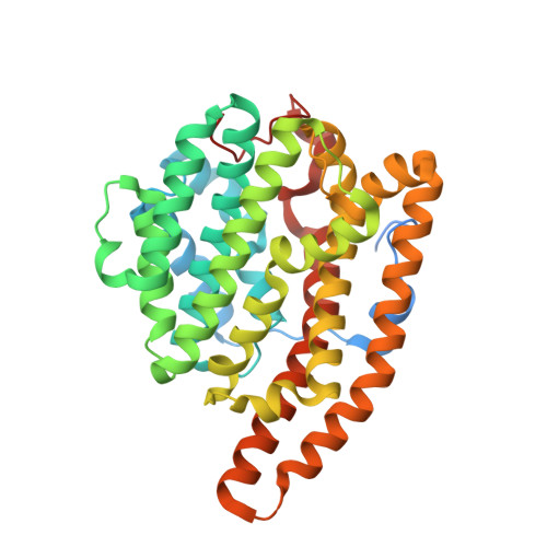
7KJG

F96M epi-isozizaene synthase: complex with 3 Mg2+ and BTAC


Experimental Data Snapshot

  • Method: X-RAY DIFFRACTION
  • Resolution: 1.30 Å
  • R-Value Free: 0.197 
  • R-Value Work: 0.177 
  • R-Value Observed: 0.177 

Starting Model: experimental
View more details

wwPDB Validation   3D Report Full Report


Ligand Structure Quality Assessment 


This is version 1.2 of the entry. See complete history


Literature

An Aromatic Cluster in the Active Site of epi -Isozizaene Synthase Is an Electrostatic Toggle for Divergent Terpene Cyclization Pathways.

Ronnebaum, T.A.Gardner, S.M.Christianson, D.W.

(2020) Biochemistry 59: 4744-4754

  • DOI: https://doi.org/10.1021/acs.biochem.0c00876
  • Primary Citation of Related Structures:  
    7KJ8, 7KJ9, 7KJD, 7KJE, 7KJF, 7KJG

  • PubMed Abstract: 

    The sesquiterpene cyclase epi -isozizaene synthase (EIZS) catalyzes the cyclization of farnesyl diphosphate to form the tricyclic precursor of the antibiotic albaflavenone. The hydrophobic active site is largely defined by aromatic residues that direct a multistep reaction sequence through multiple carbocation intermediates. The previous substitution of polar residues for a key aromatic residue, F96, converts EIZS into a high-fidelity sesquisabinene synthase: the F96S, F96M, and F96Q variants generate 78%, 91%, and 97% sesquisabinene A, respectively. Here, we report high-resolution X-ray crystal structures of two of these reprogrammed cyclases. The structures of the F96M EIZS-Mg 2+ 3 -risedronate and F96M EIZS-Mg 2+ 3 -inorganic pyrophosphate-benzyltriethylammonium cation complexes reveal structural changes in the F96 aromatic cluster that redirect the cyclization pathway leading from the bisabolyl carbocation intermediate in catalysis. The structure of the F96S EIZS-Mg 2+ 3 -neridronate complex reveals a partially occupied inhibitor and an enzyme active site caught in transition between open and closed states. Finally, three structures of wild-type EIZS complexed with the bisphosphonate inhibitors neridronate, pamidronate, and risedronate provide a foundation for understanding binding differences between wild-type and variant enzymes. These structures provide new insight regarding active site flexibility, particularly with regard to the potential for subtle expansion and contraction to accommodate ligands of varying sizes as well as bound water molecules. Additionally, these structures highlight the importance of conformational changes in the F96 aromatic cluster that could influence cation-π interactions with carbocation intermediates in catalysis.


  • Organizational Affiliation

    Roy and Diana Vagelos Laboratories, Department of Chemistry, University of Pennsylvania, Philadelphia, Pennsylvania 19104-6323, United States.


Macromolecules
Find similar proteins by:  (by identity cutoff)  |  3D Structure
Entity ID: 1
MoleculeChains Sequence LengthOrganismDetailsImage
epi-isozizaene synthase382Streptomyces coelicolorMutation(s): 1 
Gene Names: HCU77_26465
EC: 4.2.3.37
UniProt
Find proteins for Q9K499 (Streptomyces coelicolor (strain ATCC BAA-471 / A3(2) / M145))
Explore Q9K499 
Go to UniProtKB:  Q9K499
Entity Groups  
Sequence Clusters30% Identity50% Identity70% Identity90% Identity95% Identity100% Identity
UniProt GroupQ9K499
Sequence Annotations
Expand
  • Reference Sequence
Small Molecules
Ligands 5 Unique
IDChains Name / Formula / InChI Key2D Diagram3D Interactions
BTM (Subject of Investigation/LOI)
Query on BTM

Download Ideal Coordinates CCD File 
G [auth A]N-benzyl-N,N-diethylethanaminium
C13 H22 N
VBQDSLGFSUGBBE-UHFFFAOYSA-N
POP
Query on POP

Download Ideal Coordinates CCD File 
E [auth A]PYROPHOSPHATE 2-
H2 O7 P2
XPPKVPWEQAFLFU-UHFFFAOYSA-L
SO4
Query on SO4

Download Ideal Coordinates CCD File 
F [auth A]SULFATE ION
O4 S
QAOWNCQODCNURD-UHFFFAOYSA-L
GOL
Query on GOL

Download Ideal Coordinates CCD File 
H [auth A]GLYCEROL
C3 H8 O3
PEDCQBHIVMGVHV-UHFFFAOYSA-N
MG
Query on MG

Download Ideal Coordinates CCD File 
B [auth A],
C [auth A],
D [auth A]
MAGNESIUM ION
Mg
JLVVSXFLKOJNIY-UHFFFAOYSA-N
Experimental Data & Validation

Experimental Data

  • Method: X-RAY DIFFRACTION
  • Resolution: 1.30 Å
  • R-Value Free: 0.197 
  • R-Value Work: 0.177 
  • R-Value Observed: 0.177 
  • Space Group: P 1 21 1
Unit Cell:
Length ( Å )Angle ( ˚ )
a = 51.62α = 90
b = 46.95β = 97.81
c = 75.12γ = 90
Software Package:
Software NamePurpose
PHENIXrefinement
PHENIXrefinement
iMOSFLMdata reduction
Aimlessdata scaling
PHASERphasing

Structure Validation

View Full Validation Report


Ligand Structure Quality Assessment 


Entry History & Funding Information

Deposition Data


Funding OrganizationLocationGrant Number
National Institutes of Health/National Institute of General Medical Sciences (NIH/NIGMS)United StatesGM56838

Revision History  (Full details and data files)

  • Version 1.0: 2020-12-16
    Type: Initial release
  • Version 1.1: 2020-12-30
    Changes: Database references
  • Version 1.2: 2023-10-18
    Changes: Data collection, Database references, Refinement description