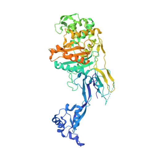
7O49

Crystal structure of Penicillin-Binding Protein 1 (PBP1) from Staphylococcus aureus


Experimental Data Snapshot

  • Method: X-RAY DIFFRACTION
  • Resolution: 3.03 Å
  • R-Value Free: 0.246 
  • R-Value Work: 0.211 
  • R-Value Observed: 0.213 

Starting Model: experimental
View more details

wwPDB Validation   3D Report Full Report


This is version 1.1 of the entry. See complete history


Literature

Integrative structural biology of the penicillin-binding protein-1 from Staphylococcus aureus , an essential component of the divisome machinery.

Martinez-Caballero, S.Mahasenan, K.V.Kim, C.Molina, R.Feltzer, R.Lee, M.Bouley, R.Hesek, D.Fisher, J.F.Munoz, I.G.Chang, M.Mobashery, S.Hermoso, J.A.

(2021) Comput Struct Biotechnol J 19: 5392-5405

  • DOI: https://doi.org/10.1016/j.csbj.2021.09.018
  • Primary Citation of Related Structures:  
    7O49, 7O4A, 7O4B, 7O4C, 7OK9

  • PubMed Abstract: 

    The penicillin-binding proteins are the enzyme catalysts of the critical transpeptidation crosslinking polymerization reaction of bacterial peptidoglycan synthesis and the molecular targets of the penicillin antibiotics. Here, we report a combined crystallographic, small-angle X-ray scattering (SAXS) in-solution structure, computational and biophysical analysis of PBP1 of  Staphylococcus aureus (sa PBP1), providing mechanistic clues about its function and regulation during cell division. The structure reveals the pedestal domain, the transpeptidase domain, and most of the linker connecting to the "penicillin-binding protein and serine/threonine kinase associated" (PASTA) domains, but not its two PASTA domains, despite their presence in the construct. To address this absence, the structure of the PASTA domains was determined at 1.5 Å resolution. Extensive molecular-dynamics simulations interpret the PASTA domains of  sa PBP1 as conformationally mobile and separated from the transpeptidase domain. This conclusion was confirmed by SAXS experiments on the full-length protein in solution. A series of crystallographic complexes with β-lactam antibiotics (as inhibitors) and penta-Gly (as a substrate mimetic) allowed the molecular characterization of both inhibition by antibiotics and binding for the donor and acceptor peptidoglycan strands. Mass-spectrometry experiments with synthetic peptidoglycan fragments revealed binding by PASTA domains in coordination with the remaining domains. The observed mobility of the PASTA domain in  sa PBP1 could play a crucial role for  in vivo  interaction with its glycosyltransferase partner in the membrane or with other components of the divisome machinery, as well as for coordination of transpeptidation and polymerization processes in the bacterial divisome.


  • Organizational Affiliation

    Department of Crystallography and Structural Biology, Institute of Physical Chemistry "Rocasolano", CSIC, 28006 Madrid, Spain.


Macromolecules
Find similar proteins by:  (by identity cutoff)  |  3D Structure
Entity ID: 1
MoleculeChains Sequence LengthOrganismDetailsImage
Penicillin-binding protein 1
A, B, C, D, E
A, B, C, D, E, F, G, H, I, J, K, L
650Staphylococcus aureus subsp. aureus COLMutation(s): 0 
Gene Names: pbp1SACOL1194
UniProt
Find proteins for A0A0H2WVW5 (Staphylococcus aureus (strain COL))
Explore A0A0H2WVW5 
Go to UniProtKB:  A0A0H2WVW5
Entity Groups  
Sequence Clusters30% Identity50% Identity70% Identity90% Identity95% Identity100% Identity
UniProt GroupA0A0H2WVW5
Sequence Annotations
Expand
  • Reference Sequence
Small Molecules
Ligands 3 Unique
IDChains Name / Formula / InChI Key2D Diagram3D Interactions
EPE
Query on EPE

Download Ideal Coordinates CCD File 
CA [auth G]
EA [auth H]
HA [auth I]
JA [auth J]
M [auth A]
CA [auth G],
EA [auth H],
HA [auth I],
JA [auth J],
M [auth A],
O [auth B],
Q [auth C],
T [auth D],
W [auth E],
Z [auth F]
4-(2-HYDROXYETHYL)-1-PIPERAZINE ETHANESULFONIC ACID
C8 H18 N2 O4 S
JKMHFZQWWAIEOD-UHFFFAOYSA-N
CD
Query on CD

Download Ideal Coordinates CCD File 
BA [auth F]
DA [auth G]
GA [auth H]
IA [auth I]
LA [auth J]
BA [auth F],
DA [auth G],
GA [auth H],
IA [auth I],
LA [auth J],
N [auth A],
P [auth B],
S [auth C],
V [auth D],
Y [auth E]
CADMIUM ION
Cd
WLZRMCYVCSSEQC-UHFFFAOYSA-N
CL
Query on CL

Download Ideal Coordinates CCD File 
AA [auth F]
FA [auth H]
KA [auth J]
MA [auth K]
NA [auth L]
AA [auth F],
FA [auth H],
KA [auth J],
MA [auth K],
NA [auth L],
R [auth C],
U [auth D],
X [auth E]
CHLORIDE ION
Cl
VEXZGXHMUGYJMC-UHFFFAOYSA-M
Experimental Data & Validation

Experimental Data

  • Method: X-RAY DIFFRACTION
  • Resolution: 3.03 Å
  • R-Value Free: 0.246 
  • R-Value Work: 0.211 
  • R-Value Observed: 0.213 
  • Space Group: P 21 21 2
Unit Cell:
Length ( Å )Angle ( ˚ )
a = 311.862α = 90
b = 197.148β = 90
c = 221.596γ = 90
Software Package:
Software NamePurpose
REFMACrefinement
PDB_EXTRACTdata extraction
XDSdata reduction
STARANISOdata scaling
PHASERphasing

Structure Validation

View Full Validation Report


Entry History 

Deposition Data

Revision History  (Full details and data files)

  • Version 1.0: 2021-11-03
    Type: Initial release
  • Version 1.1: 2024-01-31
    Changes: Data collection, Refinement description