7P9N

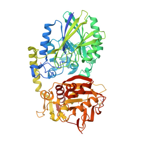
Crystal structure of CD73 in complex with AMP in the open form


Experimental Data Snapshot

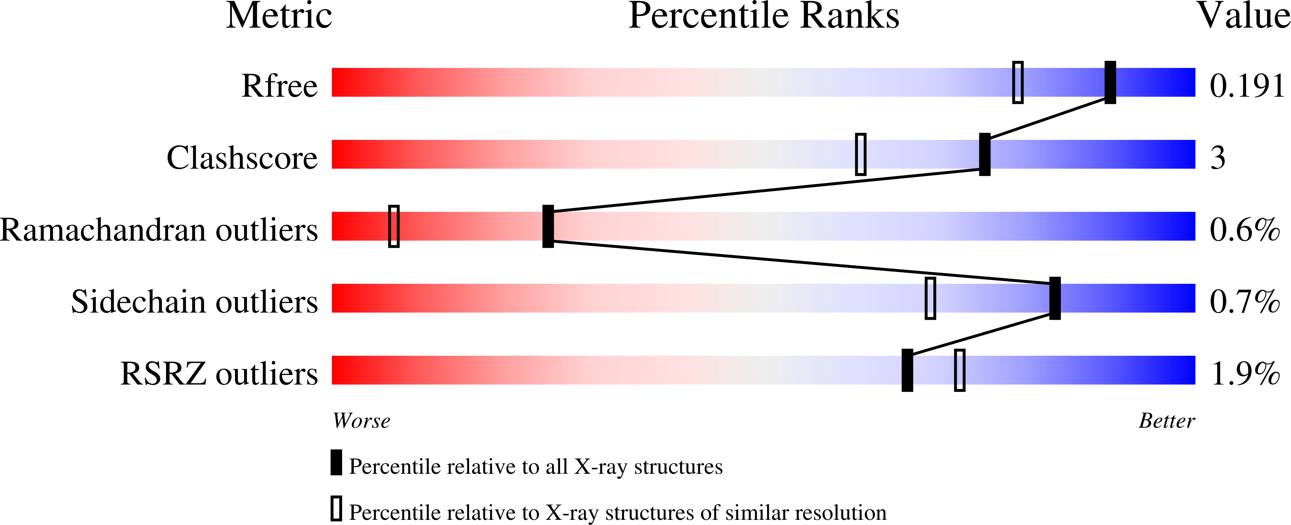
  • Method: X-RAY DIFFRACTION
  • Resolution: 1.55 Å
  • R-Value Free: 0.177 
  • R-Value Work: 0.152 
  • R-Value Observed: 0.153 

Starting Model: experimental
View more details

wwPDB Validation   3D Report Full Report


Ligand Structure Quality Assessment 


This is version 1.3 of the entry. See complete history


Literature

Substrate binding modes of purine and pyrimidine nucleotides to human ecto-5'-nucleotidase (CD73) and inhibition by their bisphosphonic acid derivatives.

Scaletti, E.Huschmann, F.U.Mueller, U.Weiss, M.S.Strater, N.

(2021) Purinergic Signal 17: 693-704

  • DOI: https://doi.org/10.1007/s11302-021-09802-w
  • Primary Citation of Related Structures:  
    7P9N, 7P9R, 7P9T, 7PA4, 7PB5, 7PBA, 7PBB, 7PBY, 7PCP, 7PD9

  • PubMed Abstract: 

    Human ecto-5-nucleotidase (CD73) is involved in purinergic signalling, which influences a diverse range of biological processes. CD73 hydrolyses AMP and is the major control point for the levels of extracellular adenosine. Inhibitors of CD73 thus block the immunosuppressive action of adenosine, a promising approach for cancer immunotherapy. Interestingly, ADP and ATP are competitive inhibitors of CD73, with the most potent small-molecule inhibitors to date being non-hydrolysable ADP analogues. While AMP is the major substrate of the enzyme, CD73 has been reported to hydrolyse other 5'-nucleoside monophosphates. Based on a fragment screening campaign at the BESSY II synchrotron, we present the binding modes of various deoxyribo- and ribonucleoside monophosphates and of four additional fragments binding to the nucleoside binding site of the open form of the enzyme. Kinetic analysis of monophosphate hydrolysis shows that ribonucleotide substrates are favoured over their deoxyribose equivalents with AMP being the best substrate. We characterised the initial step of AMP hydrolysis, the binding mode of AMP to the open conformation of CD73 and compared that to other monophosphate substrates. In addition, the inhibitory activity of various bisphosphonic acid derivatives of nucleoside diphosphates was determined. Although AMPCP remains the most potent inhibitor, replacement of the adenine base with other purines or with pyrimidines increases the K i value only between twofold and sixfold. On the other hand, these nucleobases offer new opportunities to attach substituents for improved pharmacological properties.


  • Organizational Affiliation

    Institute of Bioanalytical Chemistry, Centre for Biotechnology and Biomedicine, Leipzig University, Deutscher Platz 5, 04103, Leipzig, Germany.


Macromolecules
Find similar proteins by:  (by identity cutoff)  |  3D Structure
Entity ID: 1
MoleculeChains Sequence LengthOrganismDetailsImage
5'-nucleotidase546Homo sapiensMutation(s): 8 
Gene Names: NT5ENT5NTE
EC: 3.1.3.5 (PDB Primary Data), 3.1.3.99 (UniProt), 3.1.3.89 (UniProt), 3.1.3.35 (UniProt), 3.1.3.91 (UniProt)
UniProt & NIH Common Fund Data Resources
Find proteins for P21589 (Homo sapiens)
Explore P21589 
Go to UniProtKB:  P21589
PHAROS:  P21589
GTEx:  ENSG00000135318 
Entity Groups  
Sequence Clusters30% Identity50% Identity70% Identity90% Identity95% Identity100% Identity
UniProt GroupP21589
Sequence Annotations
Expand
  • Reference Sequence
Small Molecules
Ligands 4 Unique
IDChains Name / Formula / InChI Key2D Diagram3D Interactions
AMP (Subject of Investigation/LOI)
Query on AMP

Download Ideal Coordinates CCD File 
F [auth A]ADENOSINE MONOPHOSPHATE
C10 H14 N5 O7 P
UDMBCSSLTHHNCD-KQYNXXCUSA-N
GOL
Query on GOL

Download Ideal Coordinates CCD File 
E [auth A]GLYCEROL
C3 H8 O3
PEDCQBHIVMGVHV-UHFFFAOYSA-N
ZN
Query on ZN

Download Ideal Coordinates CCD File 
B [auth A],
C [auth A]
ZINC ION
Zn
PTFCDOFLOPIGGS-UHFFFAOYSA-N
CA
Query on CA

Download Ideal Coordinates CCD File 
D [auth A]CALCIUM ION
Ca
BHPQYMZQTOCNFJ-UHFFFAOYSA-N
Experimental Data & Validation

Experimental Data

  • Method: X-RAY DIFFRACTION
  • Resolution: 1.55 Å
  • R-Value Free: 0.177 
  • R-Value Work: 0.152 
  • R-Value Observed: 0.153 
  • Space Group: P 21 21 2
Unit Cell:
Length ( Å )Angle ( ˚ )
a = 67.345α = 90
b = 131.346β = 90
c = 66.253γ = 90
Software Package:
Software NamePurpose
REFMACrefinement
Aimlessdata scaling
PDB_EXTRACTdata extraction
XDSdata reduction
REFMACphasing

Structure Validation

View Full Validation Report


Ligand Structure Quality Assessment 


Entry History 

Deposition Data

Revision History  (Full details and data files)

  • Version 1.0: 2021-10-13
    Type: Initial release
  • Version 1.1: 2021-12-29
    Changes: Database references
  • Version 1.2: 2024-01-31
    Changes: Data collection, Refinement description
  • Version 1.3: 2024-11-13
    Changes: Structure summary