7PC1

DNA binding domain of partition protein StbA of plasmid R388


Experimental Data Snapshot

  • Method: X-RAY DIFFRACTION
  • Resolution: 1.90 Å
  • R-Value Free: 0.222 
  • R-Value Work: 0.200 
  • R-Value Observed: 0.203 

wwPDB Validation   3D Report Full Report


This is version 1.0 of the entry. See complete history


Literature

Characterization of the DNA Binding Domain of StbA, A Key Protein of A New Type of DNA Segregation System.

Quebre, V.Del Campo, I.Cuevas, A.Siguier, P.Rech, J.Le, P.T.N.Ton-Hoang, B.Cornet, F.Bouet, J.Y.Moncalian, G.de la Cruz, F.Guynet, C.

(2022) J Mol Biol 434: 167752-167752

  • DOI: https://doi.org/10.1016/j.jmb.2022.167752
  • Primary Citation of Related Structures:  
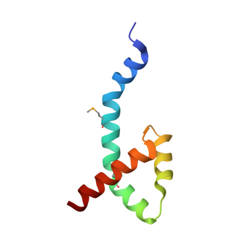
    7PC1

  • PubMed Abstract: 
  • Low-copy-number plasmids require sophisticated genetic devices to achieve efficient segregation of plasmid copies during cell division. Plasmid R388 uses a unique segregation mechanism, based on StbA, a small multifunctional protein. StbA is the key protein in a segregation system not involving a plasmid-encoded NTPase partner, it regulates the expression of several plasmid operons, and it is the main regulator of plasmid conjugation ...

    Low-copy-number plasmids require sophisticated genetic devices to achieve efficient segregation of plasmid copies during cell division. Plasmid R388 uses a unique segregation mechanism, based on StbA, a small multifunctional protein. StbA is the key protein in a segregation system not involving a plasmid-encoded NTPase partner, it regulates the expression of several plasmid operons, and it is the main regulator of plasmid conjugation. The mechanisms by which StbA, together with the centromere-like sequence stbS, achieves segregation, is largely uncharacterized. To better understand the molecular basis of R388 segregation, we determined the crystal structure of the conserved N-terminal domain of StbA to 1.9 Å resolution. It folds into an HTH DNA-binding domain, structurally related to that of the PadR subfamily II of transcriptional regulators. StbA is organized in two domains. Its N-terminal domain carries the specific stbS DNA binding activity. A truncated version of StbA, deleted of its C-terminal domain, displays only partial activities in vivo, indicating that the non-conserved C-terminal domain is required for efficient segregation and subcellular plasmid positioning. The structure of StbA DNA-binding domain also provides some insight into how StbA monomers cooperate to repress transcription by binding to the stbDR and to form the segregation complex with stbS.


    Organizational Affiliation

    Laboratoire de Microbiologie et de Génétique Moléculaires, Centre de Biologie Intégrative (CBI), Centre National de la Recherche Scientifique, Université de Toulouse, UPS, F-31000 Toulouse, France. Electronic address: Catherine.Guynet@univ-tlse3.fr.


Macromolecules
Find similar proteins by:  (by identity cutoff)  |  3D Structure
Entity ID: 1
MoleculeChainsSequence LengthOrganismDetailsImage
StbA72Escherichia coli K-12Mutation(s): 0 
UniProt
Find proteins for Q6I6C4 (Escherichia coli)
Explore Q6I6C4 
Go to UniProtKB:  Q6I6C4
Entity Groups  
Sequence Clusters30% Identity50% Identity70% Identity90% Identity95% Identity100% Identity
UniProt GroupQ6I6C4
Protein Feature View
Expand
  • Reference Sequence
Small Molecules
Modified Residues  1 Unique
IDChainsTypeFormula2D DiagramParent
MSE
Query on MSE
A
L-PEPTIDE LINKINGC5 H11 N O2 SeMET
Experimental Data & Validation

Experimental Data

  • Method: X-RAY DIFFRACTION
  • Resolution: 1.90 Å
  • R-Value Free: 0.222 
  • R-Value Work: 0.200 
  • R-Value Observed: 0.203 
  • Space Group: P 31
Unit Cell:
Length ( Å )Angle ( ˚ )
a = 45.623α = 90
b = 45.623β = 90
c = 34.393γ = 120
Software Package:
Software NamePurpose
XDSdata reduction
SCALAdata scaling
PHENIXphasing
PHENIXrefinement

Structure Validation

View Full Validation Report


Entry History & Funding Information

Deposition Data


Funding OrganizationLocationGrant Number
Spanish Ministry of Science, Innovation, and UniversitiesSpainPGC2018-093885-B-I00

Revision History  (Full details and data files)

  • Version 1.0: 2022-08-10
    Type: Initial release