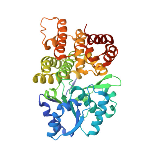
7R0P

CRYSTAL STRUCTURE OF E.coli ALCOHOL DEHYDROGENASE - FucO MUTANT F254I COMPLEXED WITH FE, NAD+, AND ETHYLENE GLYCOL


Experimental Data Snapshot

  • Method: X-RAY DIFFRACTION
  • Resolution: 1.60 Å
  • R-Value Free: 0.177 
  • R-Value Work: 0.156 

Starting Model: experimental
View more details

wwPDB Validation   3D Report Full Report


Ligand Structure Quality Assessment 


This is version 1.2 of the entry. See complete history


Literature

Structures of lactaldehyde reductase, FucO, link enzyme activity to hydrogen bond networks and conformational dynamics.

Zavarise, A.Sridhar, S.Kiema, T.R.Wierenga, R.K.Widersten, M.

(2023) FEBS J 290: 465-481

  • DOI: https://doi.org/10.1111/febs.16603
  • Primary Citation of Related Structures:  
    7QNF, 7QNI, 7QNJ, 7R0P, 7R3D, 7R5T

  • PubMed Abstract: 

    A group-III iron containing 1,2-propanediol oxidoreductase, FucO, (also known as lactaldehyde reductase) from Escherichia coli was examined regarding its structure-dynamics-function relationships in the catalysis of the NADH-dependent reduction of (2S)-lactaldehyde. Crystal structures of FucO variants in the presence or absence of cofactors have been determined, illustrating large domain movements between the apo and holo enzyme structures. Different structures of FucO variants co-crystallized with NAD + or NADH together with substrate further suggest dynamic properties of the nicotinamide moiety of the coenzyme that are important for the reaction mechanism. Modelling of the native substrate (2S)-lactaldehyde into the active site can explain the stereoselectivity exhibited by the enzyme, with a critical hydrogen bond interaction between the (2S)-hydroxyl and the side-chain of N151, as well as the previously experimentally demonstrated pro-(R) selectivity in hydride transfer from NADH to the aldehydic carbon. Furthermore, the deuterium kinetic isotope effect of hydride transfer suggests that reduction chemistry is the main rate-limiting step for turnover which is not the case in FucO catalysed alcohol oxidation. We further propose that a water molecule in the active site - hydrogen bonded to a conserved histidine (H267) and the 2'-hydroxyl of the coenzyme ribose - functions as a catalytic proton donor in the protonation of the product alcohol. A hydrogen bond network of water molecules and the side-chains of amino acid residues D360 and H267 links bulk solvent to this proposed catalytic water molecule.


  • Organizational Affiliation

    Department of Chemistry - BMC, Uppsala University, Sweden.


Macromolecules
Find similar proteins by:  (by identity cutoff)  |  3D Structure
Entity ID: 1
MoleculeChains Sequence LengthOrganismDetailsImage
Lactaldehyde reductaseA [auth AAA],
B [auth BBB]
390Escherichia coli str. K-12 substr. MG1655Mutation(s): 1 
Gene Names: fucOZ4116ECs3659
EC: 1.1.1.77
UniProt
Find proteins for P0A9S1 (Escherichia coli (strain K12))
Explore P0A9S1 
Go to UniProtKB:  P0A9S1
Entity Groups  
Sequence Clusters30% Identity50% Identity70% Identity90% Identity95% Identity100% Identity
UniProt GroupP0A9S1
Sequence Annotations
Expand
  • Reference Sequence
Small Molecules
Ligands 3 Unique
IDChains Name / Formula / InChI Key2D Diagram3D Interactions
NAD (Subject of Investigation/LOI)
Query on NAD

Download Ideal Coordinates CCD File 
C [auth AAA],
M [auth BBB]
NICOTINAMIDE-ADENINE-DINUCLEOTIDE
C21 H27 N7 O14 P2
BAWFJGJZGIEFAR-NNYOXOHSSA-N
EDO (Subject of Investigation/LOI)
Query on EDO

Download Ideal Coordinates CCD File 
D [auth AAA]
E [auth AAA]
F [auth AAA]
H [auth AAA]
I [auth AAA]
D [auth AAA],
E [auth AAA],
F [auth AAA],
H [auth AAA],
I [auth AAA],
J [auth AAA],
K [auth AAA],
L [auth AAA],
N [auth BBB],
O [auth BBB],
P [auth BBB],
Q [auth BBB],
R [auth BBB],
S [auth BBB],
U [auth BBB],
V [auth BBB],
W [auth BBB]
1,2-ETHANEDIOL
C2 H6 O2
LYCAIKOWRPUZTN-UHFFFAOYSA-N
FE (Subject of Investigation/LOI)
Query on FE

Download Ideal Coordinates CCD File 
G [auth AAA],
T [auth BBB]
FE (III) ION
Fe
VTLYFUHAOXGGBS-UHFFFAOYSA-N
Experimental Data & Validation

Experimental Data

  • Method: X-RAY DIFFRACTION
  • Resolution: 1.60 Å
  • R-Value Free: 0.177 
  • R-Value Work: 0.156 
  • Space Group: P 21 21 21
Unit Cell:
Length ( Å )Angle ( ˚ )
a = 57.98α = 90
b = 86.161β = 90
c = 147.652γ = 90
Software Package:
Software NamePurpose
REFMACrefinement
DIALSdata reduction
Aimlessdata scaling
REFMACphasing

Structure Validation

View Full Validation Report


Ligand Structure Quality Assessment 


Entry History & Funding Information

Deposition Data


Funding OrganizationLocationGrant Number
Other governmentSwedenOlle Engkvist Byggmastare Foundation - 194-0638

Revision History  (Full details and data files)

  • Version 1.0: 2022-10-19
    Type: Initial release
  • Version 1.1: 2023-02-01
    Changes: Database references
  • Version 1.2: 2024-02-07
    Changes: Data collection, Refinement description