


Structure of VspTPI - Verrucomicrobium spinosum triosephoshate isomerase
Vickers, C.J., Fraga, D., Patrick, W.M.To be published.
Experimental Data Snapshot
Starting Model: experimental
View more details
wwPDB Validation 3D Report Full Report

Entity ID: 1 | |||||
|---|---|---|---|---|---|
| Molecule | Chains | Sequence Length | Organism | Details | Image |
| Triosephosphate isomerase | 271 | Verrucomicrobium spinosum | Mutation(s): 0 EC: 5.3.1.1 | 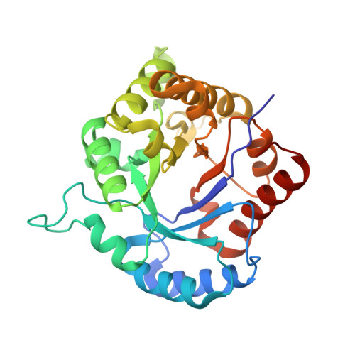 | |
UniProt | |||||
Find proteins for A0A979HMP0 (Verrucomicrobium spinosum) Explore A0A979HMP0 Go to UniProtKB: A0A979HMP0 | |||||
Entity Groups | |||||
| Sequence Clusters | 30% Identity50% Identity70% Identity90% Identity95% Identity100% Identity | ||||
| UniProt Group | A0A979HMP0 | ||||
Sequence AnnotationsExpand | |||||
| |||||
| Length ( Å ) | Angle ( ˚ ) |
|---|---|
| a = 48.99 | α = 90 |
| b = 64.118 | β = 100.787 |
| c = 94.215 | γ = 90 |
| Software Name | Purpose |
|---|---|
| PHENIX | refinement |
| MOSFLM | data reduction |
| Aimless | data scaling |
| PHASER | phasing |
| Funding Organization | Location | Grant Number |
|---|---|---|
| Royal Society of New Zealand | New Zealand | -- |


