7WJ2

Crystal structure of HLA-C*1402 complexed with 8-mer HIV gag peptide


Experimental Data Snapshot

  • Method: X-RAY DIFFRACTION
  • Resolution: 1.28 Å
  • R-Value Free: 0.178 
  • R-Value Work: 0.156 
  • R-Value Observed: 0.157 

wwPDB Validation   3D Report Full Report


This is version 1.2 of the entry. See complete history


Literature

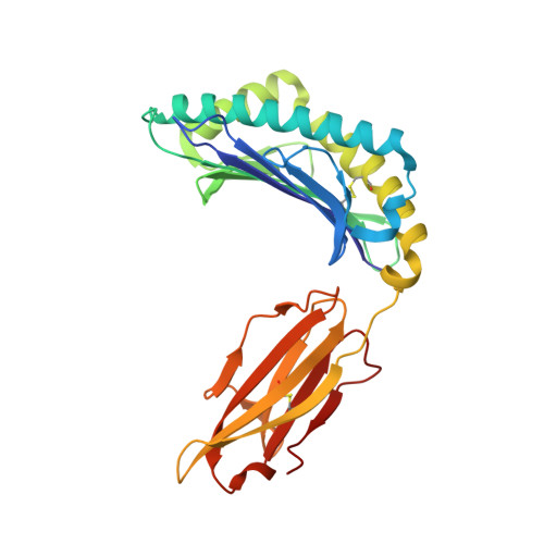
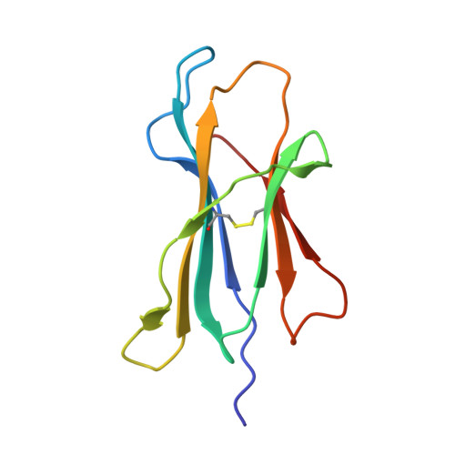
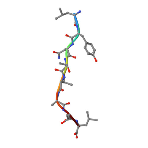
Crystal structures of N-myristoylated lipopeptide-bound HLA class I complexes indicate reorganization of B-pocket architecture upon ligand binding.

Asa, M.Morita, D.Kuroha, J.Mizutani, T.Mori, N.Mikami, B.Sugita, M.

(2022) J Biol Chem 298: 102100-102100

  • DOI: https://doi.org/10.1016/j.jbc.2022.102100
  • Primary Citation of Related Structures:  
    7WJ2, 7WJ3, 7WT3, 7WT4, 7WT5

  • PubMed Abstract: 

    Rhesus monkeys have evolved MHC-encoded class I allomorphs such as Mamu-B∗098 that are capable of binding N-myristoylated short lipopeptides rather than conventional long peptides; however, it remains unknown whether such antigen-binding molecules exist in other species, including humans. We herein demonstrate that human leukocyte antigen (HLA)-A∗24:02 and HLA-C∗14:02 proteins, which are known to bind conventional long peptides, also have the potential to bind N-myristoylated short lipopeptides. These HLA class I molecules shared a serine at position 9 (Ser9) with Mamu-B∗098, in contrast to most MHC class I molecules that harbor a larger amino acid residue, such as tyrosine, at this position. High resolution X-ray crystallographic analyses of lipopeptide-bound HLA-A∗24:02 and HLA-C∗14:02 complexes indicated that Ser9 was at the bottom of the B pocket with its small hydroxymethyl side chain directed away from the B-pocket cavity, thereby contributing to the formation of a deep hydrophobic cavity suitable for accommodating the long-chain fatty acid moiety of lipopeptide ligands. Upon peptide binding, however, we found the hydrogen-bond network involving Ser9 was reorganized, and the remodeled B pocket was able to capture the second amino acid residue (P2) of peptide ligands. Apart from the B pocket, virtually no marked alterations were observed for the A and F pockets upon peptide and lipopeptide binding. Thus, we concluded that the structural flexibility of the large B pocket of HLA-A∗2402 and HLA-C∗1402 primarily accounted for their previously unrecognized capacity to bind such chemically distinct ligands as conventional peptides and N-myristoylated lipopeptides.


  • Organizational Affiliation

    Laboratory of Cell Regulation, Institute for Life and Medical Sciences, Kyoto University, Kyoto, Japan; Laboratory of Cell Regulation and Molecular Network, Graduate School of Biostudies, Kyoto University, Kyoto, Japan.


Macromolecules
Find similar proteins by:  (by identity cutoff)  |  3D Structure
Entity ID: 1
MoleculeChains Sequence LengthOrganismDetailsImage
MHC class I protein277Homo sapiensMutation(s): 0 
Gene Names: HLA-C
UniProt
Find proteins for A0A858LG55 (Homo sapiens)
Explore A0A858LG55 
Go to UniProtKB:  A0A858LG55
Entity Groups  
Sequence Clusters30% Identity50% Identity70% Identity90% Identity95% Identity100% Identity
UniProt GroupA0A858LG55
Sequence Annotations
Expand
  • Reference Sequence
Find similar proteins by:  (by identity cutoff)  |  3D Structure
Entity ID: 2
MoleculeChains Sequence LengthOrganismDetailsImage
Beta-2-microglobulin100Homo sapiensMutation(s): 0 
Gene Names: B2MCDABP0092HDCMA22P
UniProt & NIH Common Fund Data Resources
Find proteins for P61769 (Homo sapiens)
Explore P61769 
Go to UniProtKB:  P61769
PHAROS:  P61769
GTEx:  ENSG00000166710 
Entity Groups  
Sequence Clusters30% Identity50% Identity70% Identity90% Identity95% Identity100% Identity
UniProt GroupP61769
Sequence Annotations
Expand
  • Reference Sequence

Find similar proteins by:  Sequence   |   3D Structure  

Entity ID: 3
MoleculeChains Sequence LengthOrganismDetailsImage
8-mer peptide8Human immunodeficiency virus 1Mutation(s): 0 
UniProt
Find proteins for P04591 (Human immunodeficiency virus type 1 group M subtype B (isolate HXB2))
Explore P04591 
Go to UniProtKB:  P04591
Entity Groups  
UniProt GroupP04591
Sequence Annotations
Expand
  • Reference Sequence
Small Molecules
Ligands 3 Unique
IDChains Name / Formula / InChI Key2D Diagram3D Interactions
EDO
Query on EDO

Download Ideal Coordinates CCD File 
D [auth A]
E [auth A]
F [auth A]
H [auth B]
I [auth B]
D [auth A],
E [auth A],
F [auth A],
H [auth B],
I [auth B],
J [auth B],
K [auth B]
1,2-ETHANEDIOL
C2 H6 O2
LYCAIKOWRPUZTN-UHFFFAOYSA-N
ACT
Query on ACT

Download Ideal Coordinates CCD File 
L [auth B]ACETATE ION
C2 H3 O2
QTBSBXVTEAMEQO-UHFFFAOYSA-M
NA
Query on NA

Download Ideal Coordinates CCD File 
G [auth A]SODIUM ION
Na
FKNQFGJONOIPTF-UHFFFAOYSA-N
Experimental Data & Validation

Experimental Data

  • Method: X-RAY DIFFRACTION
  • Resolution: 1.28 Å
  • R-Value Free: 0.178 
  • R-Value Work: 0.156 
  • R-Value Observed: 0.157 
  • Space Group: C 1 2 1
Unit Cell:
Length ( Å )Angle ( ˚ )
a = 92.655α = 90
b = 76.885β = 119.54
c = 62.053γ = 90
Software Package:
Software NamePurpose
PHENIXrefinement
HKL-2000data reduction
HKL-2000data scaling
HKL-2000data collection
MOLREPphasing

Structure Validation

View Full Validation Report


Entry History & Funding Information

Deposition Data


Funding OrganizationLocationGrant Number
Japan Society for the Promotion of Science (JSPS)Japan18K07172
Japan Society for the Promotion of Science (JSPS)Japan19H04805
Japan Society for the Promotion of Science (JSPS)Japan18K19563
Japan Society for the Promotion of Science (JSPS)Japan18H02852

Revision History  (Full details and data files)

  • Version 1.0: 2022-06-22
    Type: Initial release
  • Version 1.1: 2022-07-13
    Changes: Database references
  • Version 1.2: 2023-11-29
    Changes: Data collection, Refinement description