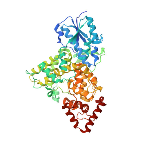
8A1H

Bacterial 6-4 photolyase from Vibrio cholerase


Experimental Data Snapshot

  • Method: X-RAY DIFFRACTION
  • Resolution: 1.65 Å
  • R-Value Free: 0.136 
  • R-Value Work: 0.119 
  • R-Value Observed: 0.119 

Starting Model: experimental
View more details

wwPDB Validation   3D Report Full Report


Ligand Structure Quality Assessment 


This is version 1.3 of the entry. See complete history


Literature

Structural and Functional Analysis of a Prokaryotic (6-4) Photolyase from the Aquatic Pathogen Vibrio Cholerae † .

Emmerich, H.J.Schneider, L.Essen, L.O.

(2023) Photochem Photobiol 99: 1248-1257

  • DOI: https://doi.org/10.1111/php.13783
  • Primary Citation of Related Structures:  
    8A1H

  • PubMed Abstract: 

    Photolyases are flavoproteins, which are able to repair UV-induced DNA lesions in a light-dependent manner. According to their substrate, they can be distinguished as CPD- and (6-4) photolyases. While CPD-photolyases repair the predominantly occurring cyclobutane pyrimidine dimer lesion, (6-4) photolyases catalyze the repair of the less prominent (6-4) photoproduct. The subgroup of prokaryotic (6-4) photolyases/FeS-BCP is one of the most ancient types of flavoproteins in the ubiquitously occurring photolyase & cryptochrome superfamily (PCSf). In contrast to canonical photolyases, prokaryotic (6-4) photolyases possess a few particular characteristics, including a lumazine derivative as antenna chromophore besides the catalytically essential flavin adenine dinucleotide as well as an elongated linker region between the N-terminal α/β-domain and the C-terminal all-α-helical domain. Furthermore, they can harbor an additional short subdomain, located at the C-terminus, with a binding site for a [4Fe-4S] cluster. So far, two crystal structures of prokaryotic (6-4) photolyases have been reported. Within this study, we present the high-resolution structure of the prokaryotic (6-4) photolyase from Vibrio cholerae and its spectroscopic characterization in terms of in vitro photoreduction and DNA-repair activity.


  • Organizational Affiliation

    Unit for Structural Biochemistry, Department of Chemistry, Philipps University Marburg, Marburg, Germany.


Macromolecules
Find similar proteins by:  (by identity cutoff)  |  3D Structure
Entity ID: 1
MoleculeChains Sequence LengthOrganismDetailsImage
6-4 photolyase (FeS-BCP, CryPro)536Vibrio choleraeMutation(s): 0 
Gene Names: VC_A0809
UniProt
Find proteins for Q9KLD7 (Vibrio cholerae serotype O1 (strain ATCC 39315 / El Tor Inaba N16961))
Explore Q9KLD7 
Go to UniProtKB:  Q9KLD7
Entity Groups  
Sequence Clusters30% Identity50% Identity70% Identity90% Identity95% Identity100% Identity
UniProt GroupQ9KLD7
Sequence Annotations
Expand
  • Reference Sequence
Small Molecules
Ligands 8 Unique
IDChains Name / Formula / InChI Key2D Diagram3D Interactions
FAD (Subject of Investigation/LOI)
Query on FAD

Download Ideal Coordinates CCD File 
B [auth A]FLAVIN-ADENINE DINUCLEOTIDE
C27 H33 N9 O15 P2
VWWQXMAJTJZDQX-UYBVJOGSSA-N
SF4
Query on SF4

Download Ideal Coordinates CCD File 
D [auth A]IRON/SULFUR CLUSTER
Fe4 S4
LJBDFODJNLIPKO-UHFFFAOYSA-N
DLZ
Query on DLZ

Download Ideal Coordinates CCD File 
C [auth A]1-deoxy-1-(6,7-dimethyl-2,4-dioxo-3,4-dihydropteridin-8(2H)-yl)-D-ribitol
C13 H18 N4 O6
SXDXRJZUAJBNFL-XKSSXDPKSA-N
EPE
Query on EPE

Download Ideal Coordinates CCD File 
E [auth A]4-(2-HYDROXYETHYL)-1-PIPERAZINE ETHANESULFONIC ACID
C8 H18 N2 O4 S
JKMHFZQWWAIEOD-UHFFFAOYSA-N
SO4
Query on SO4

Download Ideal Coordinates CCD File 
Q [auth A]
R [auth A]
S [auth A]
T [auth A]
U [auth A]
Q [auth A],
R [auth A],
S [auth A],
T [auth A],
U [auth A],
V [auth A],
W [auth A]
SULFATE ION
O4 S
QAOWNCQODCNURD-UHFFFAOYSA-L
GOL
Query on GOL

Download Ideal Coordinates CCD File 
F [auth A]
G [auth A]
H [auth A]
I [auth A]
J [auth A]
F [auth A],
G [auth A],
H [auth A],
I [auth A],
J [auth A],
K [auth A],
L [auth A],
M [auth A],
N [auth A],
O [auth A],
P [auth A]
GLYCEROL
C3 H8 O3
PEDCQBHIVMGVHV-UHFFFAOYSA-N
CL
Query on CL

Download Ideal Coordinates CCD File 
DA [auth A]CHLORIDE ION
Cl
VEXZGXHMUGYJMC-UHFFFAOYSA-M
NA
Query on NA

Download Ideal Coordinates CCD File 
AA [auth A]
BA [auth A]
CA [auth A]
X [auth A]
Y [auth A]
AA [auth A],
BA [auth A],
CA [auth A],
X [auth A],
Y [auth A],
Z [auth A]
SODIUM ION
Na
FKNQFGJONOIPTF-UHFFFAOYSA-N
Experimental Data & Validation

Experimental Data

  • Method: X-RAY DIFFRACTION
  • Resolution: 1.65 Å
  • R-Value Free: 0.136 
  • R-Value Work: 0.119 
  • R-Value Observed: 0.119 
  • Space Group: P 64 2 2
Unit Cell:
Length ( Å )Angle ( ˚ )
a = 199.687α = 90
b = 199.687β = 90
c = 76.996γ = 120
Software Package:
Software NamePurpose
PHENIXphasing
PHENIXrefinement
XDSdata reduction
Aimlessdata scaling

Structure Validation

View Full Validation Report


Ligand Structure Quality Assessment 


Entry History & Funding Information

Deposition Data


Funding OrganizationLocationGrant Number
German Research Foundation (DFG)GermanyES152/18

Revision History  (Full details and data files)

  • Version 1.0: 2023-02-08
    Type: Initial release
  • Version 1.1: 2023-02-22
    Changes: Database references
  • Version 1.2: 2023-10-04
    Changes: Data collection, Database references
  • Version 1.3: 2024-02-07
    Changes: Refinement description