8A1M

Crystal structure of the transpeptidase LdtMt2 from Mycobacterium tuberculosis in complex with maleimide analogue 4


Experimental Data Snapshot

  • Method: X-RAY DIFFRACTION
  • Resolution: 2.30 Å
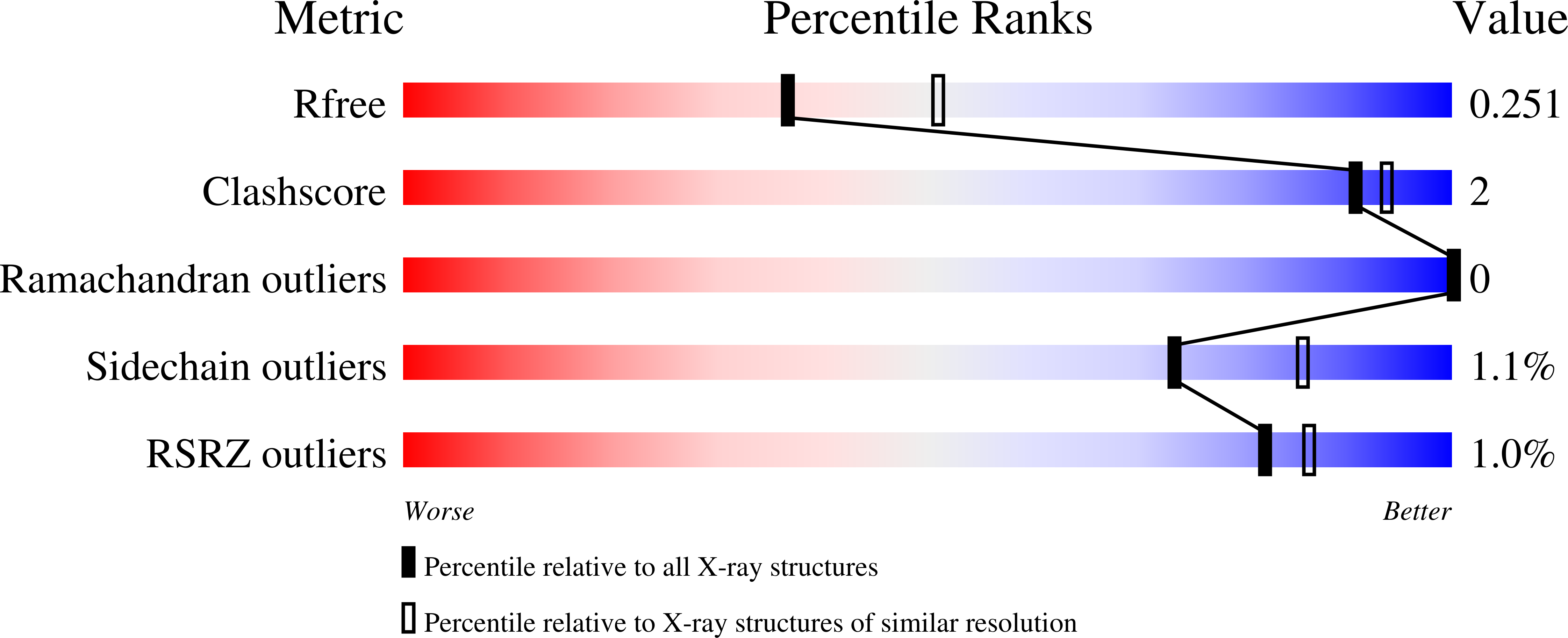
  • R-Value Free: 0.249 
  • R-Value Work: 0.218 
  • R-Value Observed: 0.220 

Starting Model: experimental
View more details

wwPDB Validation   3D Report Full Report


Ligand Structure Quality Assessment 


This is version 1.3 of the entry. See complete history


Literature

High-throughput screen with the l,d-transpeptidase Ldt Mt2 of Mycobacterium tuberculosis reveals novel classes of covalently reacting inhibitors.

de Munnik, M.Lang, P.A.De Dios Anton, F.Cacho, M.Bates, R.H.Brem, J.Rodriguez Miquel, B.Schofield, C.J.

(2023) Chem Sci 14: 7262-7278

  • DOI: https://doi.org/10.1039/d2sc06858c
  • Primary Citation of Related Structures:  
    8A1J, 8A1K, 8A1L, 8A1M, 8A1N, 8A1O, 8AHO

  • PubMed Abstract: 

    Disruption of bacterial cell wall biosynthesis in Mycobacterium tuberculosis is a promising target for treating tuberculosis. The l,d-transpeptidase Ldt Mt2 , which is responsible for the formation of 3 → 3 cross-links in the cell wall peptidoglycan, has been identified as essential for M. tuberculosis virulence. We optimised a high-throughput assay for Ldt Mt2 , and screened a targeted library of ∼10 000 electrophilic compounds. Potent inhibitor classes were identified, including established ( e.g. , β-lactams) and unexplored covalently reacting electrophilic groups ( e.g. , cyanamides). Protein-observed mass spectrometric studies reveal most classes to react covalently and irreversibly with the Ldt Mt2 catalytic cysteine (Cys354). Crystallographic analyses of seven representative inhibitors reveal induced fit involving a loop enclosing the Ldt Mt2 active site. Several of the identified compounds have a bactericidal effect on M. tuberculosis within macrophages, one with an MIC 50 value of ∼1 μM. The results provide leads for the development of new covalently reaction inhibitors of Ldt Mt2 and other nucleophilic cysteine enzymes.


  • Organizational Affiliation

    Chemistry Research Laboratory, Department of Chemistry, the Ineos Oxford Institute of Antimicrobial Research, University of Oxford 12 Mansfield Road Oxford OX1 3TA UK christopher.schofield@chem.ox.ac.uk.


Macromolecules
Find similar proteins by:  (by identity cutoff)  |  3D Structure
Entity ID: 1
MoleculeChains Sequence LengthOrganismDetailsImage
L,D-transpeptidase 2
A, B
355Mycobacterium tuberculosisMutation(s): 0 
Gene Names: ldtBMT2594V735_02606
EC: 2.3.2
UniProt
Find proteins for I6Y9J2 (Mycobacterium tuberculosis (strain ATCC 25618 / H37Rv))
Explore I6Y9J2 
Go to UniProtKB:  I6Y9J2
Entity Groups  
Sequence Clusters30% Identity50% Identity70% Identity90% Identity95% Identity100% Identity
UniProt GroupI6Y9J2
Sequence Annotations
Expand
  • Reference Sequence
Small Molecules
Ligands 6 Unique
IDChains Name / Formula / InChI Key2D Diagram3D Interactions
KUL (Subject of Investigation/LOI)
Query on KUL

Download Ideal Coordinates CCD File 
C [auth A],
P [auth B]
1-(2-fluoranyl-5-methylsulfonyl-phenyl)pyrrolidine-2,5-dione
C11 H10 F N O4 S
RISNMQBAJMCDFN-UHFFFAOYSA-N
GOL
Query on GOL

Download Ideal Coordinates CCD File 
G [auth A]GLYCEROL
C3 H8 O3
PEDCQBHIVMGVHV-UHFFFAOYSA-N
DMS
Query on DMS

Download Ideal Coordinates CCD File 
E [auth A],
F [auth A],
I [auth A],
Q [auth B],
R [auth B]
DIMETHYL SULFOXIDE
C2 H6 O S
IAZDPXIOMUYVGZ-UHFFFAOYSA-N
EDO
Query on EDO

Download Ideal Coordinates CCD File 
D [auth A],
H [auth A],
S [auth B]
1,2-ETHANEDIOL
C2 H6 O2
LYCAIKOWRPUZTN-UHFFFAOYSA-N
CL
Query on CL

Download Ideal Coordinates CCD File 
L [auth A],
M [auth A],
N [auth A],
O [auth A]
CHLORIDE ION
Cl
VEXZGXHMUGYJMC-UHFFFAOYSA-M
NA
Query on NA

Download Ideal Coordinates CCD File 
J [auth A],
K [auth A],
T [auth B]
SODIUM ION
Na
FKNQFGJONOIPTF-UHFFFAOYSA-N
Experimental Data & Validation

Experimental Data

  • Method: X-RAY DIFFRACTION
  • Resolution: 2.30 Å
  • R-Value Free: 0.249 
  • R-Value Work: 0.218 
  • R-Value Observed: 0.220 
  • Space Group: P 1 21 1
Unit Cell:
Length ( Å )Angle ( ˚ )
a = 61.7α = 90
b = 95.07β = 92.22
c = 75.45γ = 90
Software Package:
Software NamePurpose
PHENIXrefinement
XDSdata reduction
xia2data scaling
PHASERphasing
PDB_EXTRACTdata extraction

Structure Validation

View Full Validation Report


Ligand Structure Quality Assessment 


Entry History & Funding Information

Deposition Data


Funding OrganizationLocationGrant Number
Biotechnology and Biological Sciences Research Council (BBSRC)United Kingdom--

Revision History  (Full details and data files)

  • Version 1.0: 2023-06-14
    Type: Initial release
  • Version 1.1: 2023-08-02
    Changes: Database references
  • Version 1.2: 2024-02-07
    Changes: Data collection, Refinement description
  • Version 1.3: 2024-10-16
    Changes: Structure summary