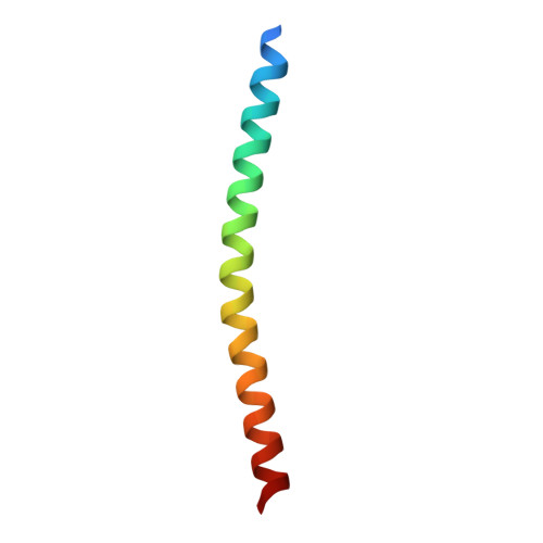
Cryo-electron microscopy of the f1 filamentous phage reveals insights into viral infection and assembly.
Conners, R., Leon-Quezada, R.I., McLaren, M., Bennett, N.J., Daum, B., Rakonjac, J., Gold, V.A.M.(2023) Nat Commun 14: 2724-2724
- PubMed: 37169795
- DOI: https://doi.org/10.1038/s41467-023-37915-w
- Primary Citation of Related Structures:
8B3O, 8B3P, 8B3Q - PubMed Abstract:
Phages are viruses that infect bacteria and dominate every ecosystem on our planet. As well as impacting microbial ecology, physiology and evolution, phages are exploited as tools in molecular biology and biotechnology. This is particularly true for the Ff (f1, fd or M13) phages, which represent a widely distributed group of filamentous viruses. Over nearly five decades, Ffs have seen an extraordinary range of applications, yet the complete structure of the phage capsid and consequently the mechanisms of infection and assembly remain largely mysterious. In this work, we use cryo-electron microscopy and a highly efficient system for production of short Ff-derived nanorods to determine a structure of a filamentous virus including the tips. We show that structure combined with mutagenesis can identify phage domains that are important in bacterial attack and for release of new progeny, allowing new models to be proposed for the phage lifecycle.
- Living Systems Institute, University of Exeter, Stocker Road, Exeter, EX4 4QD, UK.
Organizational Affiliation:


