8BE9

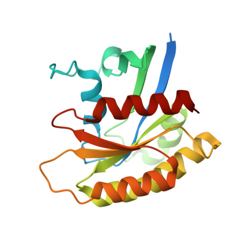
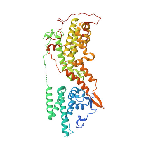
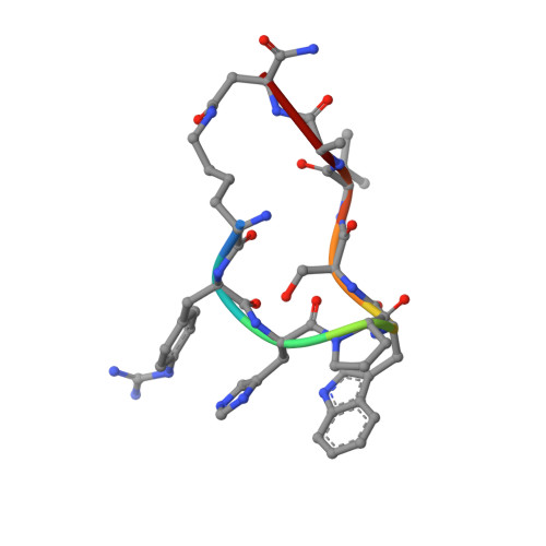
Crystal structure of SOS1-HRas-peptidomimetic5


Experimental Data Snapshot

  • Method: X-RAY DIFFRACTION
  • Resolution: 2.51 Å
  • R-Value Free: 0.229 
  • R-Value Work: 0.190 
  • R-Value Observed: 0.192 

Starting Model: experimental
View more details

wwPDB Validation   3D Report Full Report


This is version 1.2 of the entry. See complete history


Literature

Nanobody Loop Mimetics Enhance Son of Sevenless 1-Catalyzed Nucleotide Exchange on RAS.

Van Holsbeeck, K.Fischer, B.Gonzalez, S.Gadais, C.Versees, W.Martins, J.C.Martin, C.Wohlkonig, A.Steyaert, J.Ballet, S.

(2023) Angew Chem Int Ed Engl 62: e202219095-e202219095

  • DOI: https://doi.org/10.1002/anie.202219095
  • Primary Citation of Related Structures:  
    8BE6, 8BE7, 8BE8, 8BE9, 8BEA

  • PubMed Abstract: 

    RAS proteins control various intracellular signaling networks. Mutations at specific locations were shown to stabilize their active guanosine triphosphate (GTP)-bound state, which is associated with the development of multiple cancers. An attractive approach to modulate RAS signaling is through its regulatory guanine nucleotide exchange factor (GEF) son of sevenless 1 (SOS1). With the recent discovery of Nanobody14 (Nb14), which potently enhances SOS1-catalyzed nucleotide exchange on RAS, we explored the feasibility of developing peptide mimetics by structurally mimicking the complementarity-determining region 3 (CDR3). Guided by a biochemical GEF assay and X-ray co-crystal structures, successive rounds of optimization and gradual conformational rigidification led to CDR3 mimetics showing half of the maximal activation potential of Nb14 with an EC 50 value of 29 μM. Altogether, this study demonstrated that peptides able to modulate a protein-protein interaction can be obtained by structural mimicry of a Nb paratope.


  • Organizational Affiliation

    Research Group of Organic Chemistry, Vrije Universiteit Brussel, Pleinlaan 2, 1050, Brussels, Belgium.


Macromolecules
Find similar proteins by:  (by identity cutoff)  |  3D Structure
Entity ID: 1
MoleculeChains Sequence LengthOrganismDetailsImage
GTPase HRasA [auth R]186Homo sapiensMutation(s): 0 
Gene Names: HRASHRAS1
EC: 3.6.5.2
UniProt & NIH Common Fund Data Resources
Find proteins for P01112 (Homo sapiens)
Explore P01112 
Go to UniProtKB:  P01112
PHAROS:  P01112
GTEx:  ENSG00000174775 
Entity Groups  
Sequence Clusters30% Identity50% Identity70% Identity90% Identity95% Identity100% Identity
UniProt GroupP01112
Sequence Annotations
Expand
  • Reference Sequence
Find similar proteins by:  (by identity cutoff)  |  3D Structure
Entity ID: 2
MoleculeChains Sequence LengthOrganismDetailsImage
Son of sevenless homolog 1B [auth S]507Homo sapiensMutation(s): 0 
Gene Names: SOS1
UniProt & NIH Common Fund Data Resources
Find proteins for Q07889 (Homo sapiens)
Explore Q07889 
Go to UniProtKB:  Q07889
PHAROS:  Q07889
GTEx:  ENSG00000115904 
Entity Groups  
Sequence Clusters30% Identity50% Identity70% Identity90% Identity95% Identity100% Identity
UniProt GroupQ07889
Sequence Annotations
Expand
  • Reference Sequence

Find similar proteins by:  Sequence   |   3D Structure  

Entity ID: 3
MoleculeChains Sequence LengthOrganismDetailsImage
SOS1-HRas-peptidomimetic5C [auth P]9synthetic constructMutation(s): 0 
Sequence Annotations
Expand
  • Reference Sequence
Small Molecules
Ligands 2 Unique
IDChains Name / Formula / InChI Key2D Diagram3D Interactions
FMT
Query on FMT

Download Ideal Coordinates CCD File 
G [auth R]
H [auth R]
I [auth R]
J [auth R]
K [auth R]
G [auth R],
H [auth R],
I [auth R],
J [auth R],
K [auth R],
L [auth R],
M [auth S],
N [auth S],
O [auth S],
P [auth S],
Q [auth S],
R [auth S],
S,
T [auth S]
FORMIC ACID
C H2 O2
BDAGIHXWWSANSR-UHFFFAOYSA-N
CL
Query on CL

Download Ideal Coordinates CCD File 
D [auth R],
E [auth R],
F [auth R]
CHLORIDE ION
Cl
VEXZGXHMUGYJMC-UHFFFAOYSA-M
Modified Residues  2 Unique
IDChains TypeFormula2D DiagramParent
7XC
Query on 7XC
C [auth P]D-PEPTIDE LINKINGC10 H14 N4 O2PHE
XSN
Query on XSN
C [auth P]L-PEPTIDE LINKINGC4 H8 N2 O3ASN
Experimental Data & Validation

Experimental Data

  • Method: X-RAY DIFFRACTION
  • Resolution: 2.51 Å
  • R-Value Free: 0.229 
  • R-Value Work: 0.190 
  • R-Value Observed: 0.192 
  • Space Group: I 4 2 2
Unit Cell:
Length ( Å )Angle ( ˚ )
a = 150.41α = 90
b = 150.41β = 90
c = 200.98γ = 90
Software Package:
Software NamePurpose
PHENIXrefinement
XDSdata reduction
XDSdata scaling
PHENIXphasing

Structure Validation

View Full Validation Report


Entry History & Funding Information

Deposition Data


Funding OrganizationLocationGrant Number
Research Foundation - Flanders (FWO)Belgium--

Revision History  (Full details and data files)

  • Version 1.0: 2023-04-26
    Type: Initial release
  • Version 1.1: 2023-06-14
    Changes: Database references
  • Version 1.2: 2024-02-07
    Changes: Data collection, Refinement description