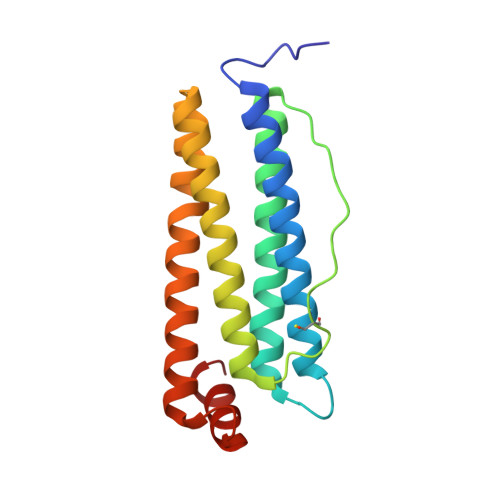
8CPV

Human apoferritin


Experimental Data Snapshot

  • Method: ELECTRON MICROSCOPY
  • Resolution: 1.76 Å
  • Aggregation State: PARTICLE 
  • Reconstruction Method: SINGLE PARTICLE 

wwPDB Validation   3D Report Full Report


This is version 1.2 of the entry. See complete history


Literature

Super-resolution fluorescence imaging of cryosamples does not limit achievable resolution in cryoEM.

Last, M.G.F.Noteborn, W.E.M.Voortman, L.M.Sharp, T.H.

(2023) J Struct Biol 215: 108040-108040

  • DOI: https://doi.org/10.1016/j.jsb.2023.108040
  • Primary Citation of Related Structures:  
    8CPM, 8CPS, 8CPT, 8CPU, 8CPV, 8CPW, 8CPX

  • PubMed Abstract: 

    Correlated super-resolution cryo-fluorescence and cryo-electron microscopy (cryoEM) has been gaining popularity as a method to investigate biological samples with high resolution and specificity. A concern in this combined method (called SR-cryoCLEM), however, is whether and how fluorescence imaging prior to cryoEM acquisition is detrimental to sample integrity. In this report, we investigated the effect of high-dose laser light (405, 488, and 561 nm) irradiation on apoferritin samples prepared for cryoEM with excitation wavelengths commonly used in fluorescence microscopy, and compared these samples to controls that were kept in the dark. We found that laser illumination, of equal duration and intensity as used in cryo-single molecule localization microscopy (cryoSMLM) and in the presence of high concentrations of fluorescent protein, did not affect the achievable resolution in cryoEM, with final reconstructions reaching resolutions of ∼ 1.8 Å regardless of the laser illumination. The finding that super-resolution fluorescence imaging of cryosamples prior to cryoEM data acquisition does not limit the achievable resolution suggests that super-resolution cryo-fluorescence microscopy and in situ structural biology using cryoEM are entirely compatible.


  • Organizational Affiliation

    Department of Cell and Chemical Biology, Leiden University Medical Centre, 2300 RC Leiden, The Netherlands.


Macromolecules
Find similar proteins by:  (by identity cutoff)  |  3D Structure
Entity ID: 1
MoleculeChains Sequence LengthOrganismDetailsImage
Ferritin heavy chain, N-terminally processed173Homo sapiensMutation(s): 1 
Gene Names: FTH1FTHFTHL6OK/SW-cl.84PIG15
UniProt & NIH Common Fund Data Resources
Find proteins for P02794 (Homo sapiens)
Explore P02794 
Go to UniProtKB:  P02794
PHAROS:  P02794
GTEx:  ENSG00000167996 
Entity Groups  
Sequence Clusters30% Identity50% Identity70% Identity90% Identity95% Identity100% Identity
UniProt GroupP02794
Sequence Annotations
Expand
  • Reference Sequence
Small Molecules
Ligands 2 Unique
IDChains Name / Formula / InChI Key2D Diagram3D Interactions
MG
Query on MG

Download Ideal Coordinates CCD File 
AA [auth 1]
CA [auth 2]
DA [auth 2]
FA [auth 4]
HA [auth 6]
AA [auth 1],
CA [auth 2],
DA [auth 2],
FA [auth 4],
HA [auth 6],
IA [auth 6],
KA [auth A],
MA [auth B],
QA [auth G],
SA [auth H],
TA [auth H],
XA [auth M],
Z [auth 1],
ZA [auth O]
MAGNESIUM ION
Mg
JLVVSXFLKOJNIY-UHFFFAOYSA-N
NA
Query on NA

Download Ideal Coordinates CCD File 
AB [auth P]
BA [auth 2]
BB [auth Q]
CB [auth S]
DB [auth U]
AB [auth P],
BA [auth 2],
BB [auth Q],
CB [auth S],
DB [auth U],
EA [auth 4],
EB [auth W],
FB [auth X],
GA [auth 6],
GB [auth Y],
HB [auth a],
IB [auth e],
JA [auth A],
JB [auth r],
LA [auth B],
NA [auth E],
OA [auth F],
PA [auth G],
RA [auth H],
UA [auth I],
VA [auth K],
WA [auth M],
Y [auth 1],
YA [auth O]
SODIUM ION
Na
FKNQFGJONOIPTF-UHFFFAOYSA-N
Modified Residues  1 Unique
IDChains TypeFormula2D DiagramParent
CSX
Query on CSX
A [auth 1]
B [auth 2]
C [auth 4]
D [auth 6]
E [auth A]
A [auth 1],
B [auth 2],
C [auth 4],
D [auth 6],
E [auth A],
F [auth B],
G [auth E],
H [auth F],
I [auth G],
J [auth H],
K [auth I],
L [auth K],
M,
N [auth O],
O [auth P],
P [auth Q],
Q [auth S],
R [auth U],
S [auth W],
T [auth X],
U [auth Y],
V [auth a],
W [auth e],
X [auth r]
L-PEPTIDE LINKINGC3 H7 N O3 SCYS
Experimental Data & Validation

Experimental Data

  • Method: ELECTRON MICROSCOPY
  • Resolution: 1.76 Å
  • Aggregation State: PARTICLE 
  • Reconstruction Method: SINGLE PARTICLE 
EM Software:
TaskSoftware PackageVersion
MODEL REFINEMENTPHENIX

Structure Validation

View Full Validation Report


Entry History & Funding Information

Deposition Data


Funding OrganizationLocationGrant Number
Netherlands Organisation for Scientific Research (NWO)NetherlandsVI.Vidi.193.014

Revision History  (Full details and data files)

  • Version 1.0: 2023-03-15
    Type: Initial release
  • Version 1.1: 2023-11-15
    Changes: Data collection, Database references, Refinement description
  • Version 1.2: 2023-11-22
    Changes: Database references