8HGW

Crystal structure of MehpH in complex with MBP


Experimental Data Snapshot

  • Method: X-RAY DIFFRACTION
  • Resolution: 2.80 Å
  • R-Value Free: 0.260 
  • R-Value Work: 0.228 
  • R-Value Observed: 0.229 

wwPDB Validation   3D Report Full Report


Ligand Structure Quality Assessment 


This is version 1.2 of the entry. See complete history


Literature

Molecular insights into the catalytic mechanism of plasticizer degradation by a monoalkyl phthalate hydrolase.

Chen, Y.Wang, Y.Xu, Y.Sun, J.Yang, L.Feng, C.Wang, J.Zhou, Y.Zhang, Z.M.Wang, Y.

(2023) Commun Chem 6: 45-45

  • DOI: https://doi.org/10.1038/s42004-023-00846-0
  • Primary Citation of Related Structures:  
    8HGV, 8HGW

  • PubMed Abstract: 

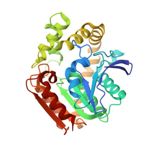
    Phthalate acid esters (PAEs), a group of xenobiotic compounds used extensively as plasticizers, have attracted increasing concern for adverse effects to human health and the environment. Microbial degradation relying on PAE hydrolases is a promising treatment. However, only a limited number of PAE hydrolases were characterized to date. Here we report the structures of MehpH, a monoalkyl phthalate (MBP) hydrolase that catalyzes the reaction of MBP to phthalic acid and the corresponding alcohol, in apo and ligand-bound form. The structures reveal a positively-charged catalytic center, complementary to the negatively-charged carboxyl group on MBP, and a penetrating tunnel that serves as exit of alcohol. The study provides a first glimpse into the enzyme-substrate binding model for PAE hydrolases, leading strong support to the development of better enzymes in the future.


  • Organizational Affiliation

    School of Biology and Biological Engineering, South China University of Technology, Guangzhou, 510006, China.


Macromolecules
Find similar proteins by:  (by identity cutoff)  |  3D Structure
Entity ID: 1
MoleculeChains Sequence LengthOrganismDetailsImage
Monoalkyl phthalate hydrolase
A, B, C, D
289Gordonia sp. P8219Mutation(s): 0 
Gene Names: mehpH
UniProt
Find proteins for Q2MHH5 (Gordonia sp. P8219)
Explore Q2MHH5 
Go to UniProtKB:  Q2MHH5
Entity Groups  
Sequence Clusters30% Identity50% Identity70% Identity90% Identity95% Identity100% Identity
UniProt GroupQ2MHH5
Sequence Annotations
Expand
  • Reference Sequence
Experimental Data & Validation

Experimental Data

  • Method: X-RAY DIFFRACTION
  • Resolution: 2.80 Å
  • R-Value Free: 0.260 
  • R-Value Work: 0.228 
  • R-Value Observed: 0.229 
  • Space Group: I 1 2 1
Unit Cell:
Length ( Å )Angle ( ˚ )
a = 76.95α = 90
b = 48.95β = 93.7297
c = 306.325γ = 90
Software Package:
Software NamePurpose
PHENIXrefinement
XDSdata reduction
XDSdata scaling
PHASERphasing
Cootmodel building

Structure Validation

View Full Validation Report


Ligand Structure Quality Assessment 


Entry History & Funding Information

Deposition Data


Funding OrganizationLocationGrant Number
Not funded--

Revision History  (Full details and data files)

  • Version 1.0: 2023-03-15
    Type: Initial release
  • Version 1.1: 2023-09-06
    Changes: Data collection, Refinement description, Source and taxonomy
  • Version 1.2: 2024-04-03
    Changes: Refinement description