8OSC

Structure of Homo sapiens 2'-deoxynucleoside 5'-phosphate N-hydrolase 1 (DNPH1) bound to deoxyuridine 5'- monophosphate


Experimental Data Snapshot

  • Method: X-RAY DIFFRACTION
  • Resolution: 1.42 Å
  • R-Value Free: 
    0.210 (Depositor), 0.210 (DCC) 
  • R-Value Work: 
    0.197 (Depositor), 0.200 (DCC) 
  • R-Value Observed: 
    0.198 (Depositor) 

Starting Model: experimental
View more details

wwPDB Validation   3D Report Full Report


Ligand Structure Quality Assessment 

Created with Raphaël 2.3.0Worse 01 BetterLigand structure goodness of fit to experimental dataBest fitted UMPClick on this verticalbar to view details

This is version 1.1 of the entry. See complete history


Literature

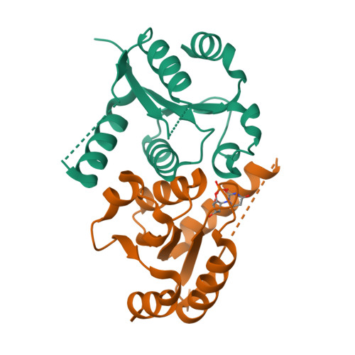
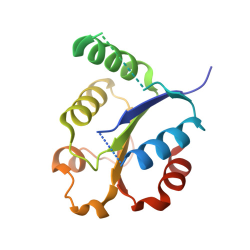
Human 2'-Deoxynucleoside 5'-Phosphate N -Hydrolase 1: Mechanism of 2'-Deoxyuridine 5'-Monophosphate Hydrolysis.

Devi, S.Carberry, A.E.Zickuhr, G.M.Dickson, A.L.Harrison, D.J.da Silva, R.G.

(2023) Biochemistry 62: 2658-2668

  • DOI: https://doi.org/10.1021/acs.biochem.3c00369
  • Primary Citation of Related Structures:  
    8OS9, 8OSC

  • PubMed Abstract: 

    The enzyme 2'-deoxynucleoside 5'-phosphate N -hydrolase 1 (DNPH1) catalyzes the N -ribosidic bond cleavage of 5-hydroxymethyl-2'-deoxyuridine 5'-monophosphate to generate 2-deoxyribose 5-phosphate and 5-hydroxymethyluracil. DNPH1 accepts other 2'-deoxynucleoside 5'-monophosphates as slow-reacting substrates. DNPH1 inhibition is a promising strategy to overcome resistance to and potentiate anticancer poly(ADP-ribose) polymerase inhibitors. We solved the crystal structure of unliganded human DNPH1 and took advantage of the slow reactivity of 2'-deoxyuridine 5'-monophosphate (dUMP) as a substrate to obtain a crystal structure of the DNPH1:dUMP Michaelis complex. In both structures, the carboxylate group of the catalytic Glu residue, proposed to act as a nucleophile in covalent catalysis, forms an apparent low-barrier hydrogen bond with the hydroxyl group of a conserved Tyr residue. The crystal structures are supported by functional data, with liquid chromatography-mass spectrometry analysis showing that DNPH1 incubation with dUMP leads to slow yet complete hydrolysis of the substrate. A direct UV-vis absorbance-based assay allowed characterization of DNPH1 kinetics at low dUMP concentrations. A bell-shaped pH-rate profile indicated that acid-base catalysis is operational and that for maximum k cat / K M , two groups with an average p K a of 6.4 must be deprotonated, while two groups with an average p K a of 8.2 must be protonated. A modestly inverse solvent viscosity effect rules out diffusional processes involved in dUMP binding to and possibly uracil release from the enzyme as rate limiting to k cat / K M . Solvent deuterium isotope effects on k cat / K M and k cat were inverse and unity, respectively. A reaction mechanism for dUMP hydrolysis is proposed.


  • Organizational Affiliation

    School of Biology, Biomedical Sciences Research Complex, University of St Andrews, St Andrews KY16 9ST, U.K.


Macromolecules
Find similar proteins by:  (by identity cutoff)  |  3D Structure
Entity ID: 1
MoleculeChains Sequence LengthOrganismDetailsImage
2'-deoxynucleoside 5'-phosphate N-hydrolase 1
A, B
145Homo sapiensMutation(s): 0 
Gene Names: DNPH1C6orf108RCL
EC: 3.2.2
UniProt & NIH Common Fund Data Resources
Find proteins for O43598 (Homo sapiens)
Explore O43598 
Go to UniProtKB:  O43598
PHAROS:  O43598
GTEx:  ENSG00000112667 
Entity Groups  
Sequence Clusters30% Identity50% Identity70% Identity90% Identity95% Identity100% Identity
UniProt GroupO43598
Sequence Annotations
Expand
  • Reference Sequence
Small Molecules
Ligands 1 Unique
IDChains Name / Formula / InChI Key2D Diagram3D Interactions
UMP (Subject of Investigation/LOI)
Query on UMP

Download Ideal Coordinates CCD File 
C [auth B]2'-DEOXYURIDINE 5'-MONOPHOSPHATE
C9 H13 N2 O8 P
JSRLJPSBLDHEIO-SHYZEUOFSA-N
Experimental Data & Validation

Experimental Data

  • Method: X-RAY DIFFRACTION
  • Resolution: 1.42 Å
  • R-Value Free:  0.210 (Depositor), 0.210 (DCC) 
  • R-Value Work:  0.197 (Depositor), 0.200 (DCC) 
  • R-Value Observed: 0.198 (Depositor) 
Space Group: C 1 2 1
Unit Cell:
Length ( Å )Angle ( ˚ )
a = 107.354α = 90
b = 38.772β = 120.29
c = 66.195γ = 90
Software Package:
Software NamePurpose
PHENIXrefinement
xia2data reduction
DIALSdata scaling
PHASERphasing

Structure Validation

View Full Validation Report


Ligand Structure Quality Assessment 

Created with Raphaël 2.3.0Worse 01 BetterLigand structure goodness of fit to experimental dataBest fitted UMPClick on this verticalbar to view details

Entry History & Funding Information

Deposition Data


Funding OrganizationLocationGrant Number
Other privateUnited KingdomIBioIC 2021-01-01

Revision History  (Full details and data files)

  • Version 1.0: 2023-08-30
    Type: Initial release
  • Version 1.1: 2023-09-13
    Changes: Database references