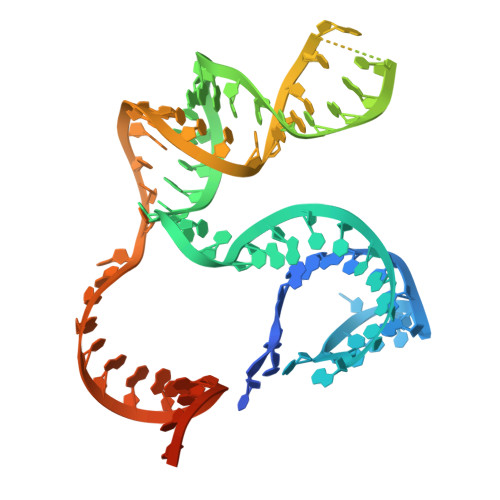
RCSB PDB - 9CEX: Spizellomyces punctatus Fanzor (SpuFz) State 4

 9CEX

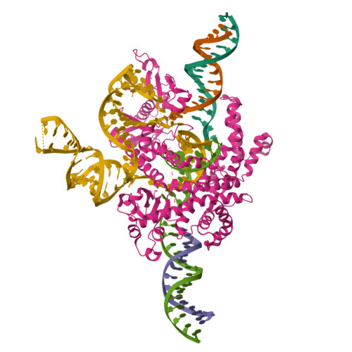
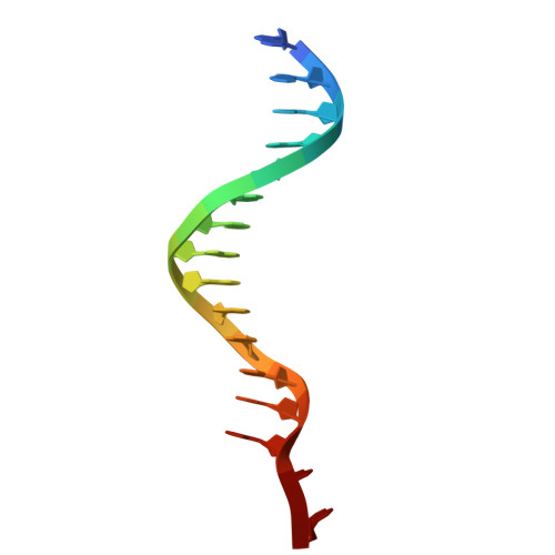
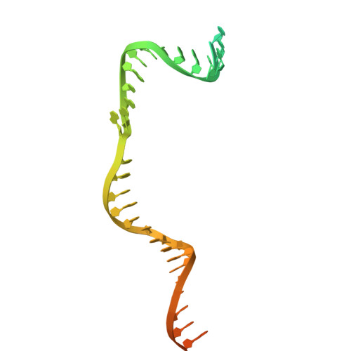
Spizellomyces punctatus Fanzor (SpuFz) State 4


Experimental Data Snapshot

  • Method: ELECTRON MICROSCOPY
  • Resolution: 3.27 Å
  • Aggregation State: PARTICLE 
  • Reconstruction Method: SINGLE PARTICLE 

wwPDB Validation   3D Report Full Report


This is version 2.1 of the entry. See complete history


Literature

Structural insights into the diversity and DNA cleavage mechanism of Fanzor.

Xu, P.Saito, M.Faure, G.Maguire, S.Chau-Duy-Tam Vo, S.Wilkinson, M.E.Kuang, H.Wang, B.Rice, W.J.Macrae, R.K.Zhang, F.

(2024) Cell 187: 5238-5252.e20

  • DOI: https://doi.org/10.1016/j.cell.2024.07.050
  • Primary Citation of Related Structures:  
    9CER, 9CES, 9CET, 9CEU, 9CEV, 9CEW, 9CEX, 9CEY, 9CEZ, 9CF0, 9CF1, 9CF2, 9CF3

  • PubMed Abstract: 

    Fanzor (Fz) is an ωRNA-guided endonuclease extensively found throughout the eukaryotic domain with unique gene editing potential. Here, we describe the structures of Fzs from three different organisms. We find that Fzs share a common ωRNA interaction interface, regardless of the length of the ωRNA, which varies considerably across species. The analysis also reveals Fz's mode of DNA recognition and unwinding capabilities as well as the presence of a non-canonical catalytic site. The structures demonstrate how protein conformations of Fz shift to allow the binding of double-stranded DNA to the active site within the R-loop. Mechanistically, examination of structures in different states shows that the conformation of the lid loop on the RuvC domain is controlled by the formation of the guide/DNA heteroduplex, regulating the activation of nuclease and DNA double-stranded displacement at the single cleavage site. Our findings clarify the mechanism of Fz, establishing a foundation for engineering efforts.


  • Organizational Affiliation

    Broad Institute of MIT and Harvard, Cambridge, MA 02142, USA; McGovern Institute for Brain Research at MIT, Cambridge, MA 02139, USA; Department of Brain and Cognitive Science, Massachusetts Institute of Technology, Cambridge, MA 02139, USA; Department of Biological Engineering, Massachusetts Institute of Technology, Cambridge, MA 02139, USA; Howard Hughes Medical Institute, Cambridge, MA 02139, USA.


Macromolecules

Find similar proteins by:  (by identity cutoff)  |  3D Structure
Entity ID: 4
MoleculeChains Sequence LengthOrganismDetailsImage
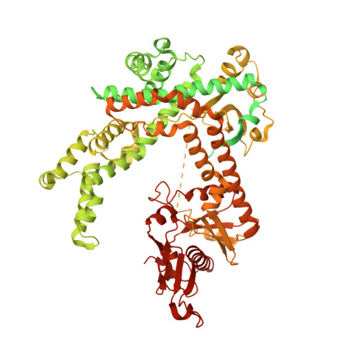
Maltose/maltodextrin-binding periplasmic protein,Spizellomyces punctatus Fanzor 1D [auth P]1,032Escherichia coli K-12Spizellomyces punctatus
This entity is chimeric
Mutation(s): 0 
Gene Names: malEb4034JW3994SPPG_07727
UniProt
Find proteins for P0AEX9 (Escherichia coli (strain K12))
Explore P0AEX9 
Go to UniProtKB:  P0AEX9
Find proteins for A0A0L0H5U9 (Spizellomyces punctatus (strain DAOM BR117))
Explore A0A0L0H5U9 
Go to UniProtKB:  A0A0L0H5U9
Entity Groups  
Sequence Clusters30% Identity50% Identity70% Identity90% Identity95% Identity100% Identity
UniProt GroupsA0A0L0H5U9P0AEX9
Sequence Annotations
Expand
  • Reference Sequence
Experimental Data & Validation

Experimental Data

  • Method: ELECTRON MICROSCOPY
  • Resolution: 3.27 Å
  • Aggregation State: PARTICLE 
  • Reconstruction Method: SINGLE PARTICLE 
EM Software:
TaskSoftware PackageVersion
MODEL REFINEMENTPHENIX1.21_5207

Structure Validation

View Full Validation Report


Entry History & Funding Information

Deposition Data


Funding OrganizationLocationGrant Number
Howard Hughes Medical Institute (HHMI)United States--

Revision History  (Full details and data files)

  • Version 1.0: 2024-09-11
    Type: Initial release
  • Version 2.0: 2024-09-25
    Type: Coordinate replacement
    Reason: Model completeness
    Changes: Advisory, Atomic model, Data collection, Database references, Derived calculations, Non-polymer description, Source and taxonomy, Structure summary
  • Version 2.1: 2024-10-02
    Changes: Data collection, Database references