9GMA | pdb_00009gma

MukBEF in a DNA capture state (dimer)


Experimental Data Snapshot

  • Method: ELECTRON MICROSCOPY
  • Resolution: 9.10 Å
  • Aggregation State: PARTICLE 
  • Reconstruction Method: SINGLE PARTICLE 

wwPDB Validation   3D Report Full Report


This is version 1.1 of the entry. See complete history


Literature

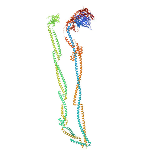
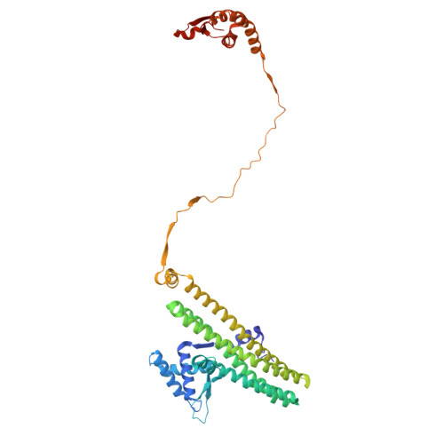
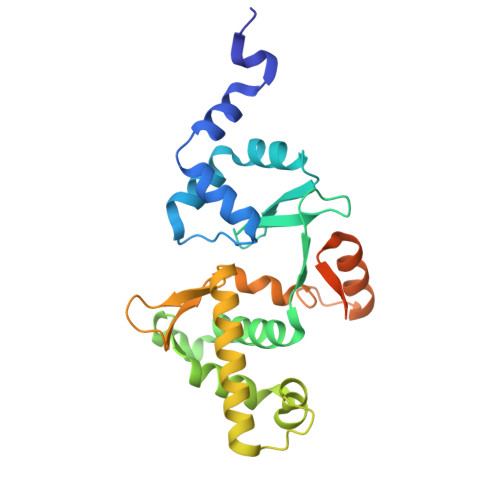
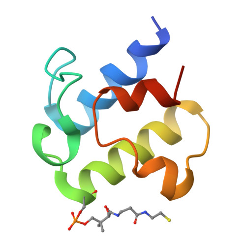
Mechanism of DNA capture by the MukBEF SMC complex and its inhibition by a viral DNA mimic.

Burmann, F.Clifton, B.Koekemoer, S.Wilkinson, O.J.Kimanius, D.Dillingham, M.S.Lowe, J.

(2025) Cell 

  • DOI: https://doi.org/10.1016/j.cell.2025.02.032
  • Primary Citation of Related Structures:  
    9GM6, 9GM7, 9GM8, 9GM9, 9GMA, 9GMB, 9GMD

  • PubMed Abstract: 

    Ring-like structural maintenance of chromosome (SMC) complexes are crucial for genome organization and operate through mechanisms of DNA entrapment and loop extrusion. Here, we explore the DNA loading process of the bacterial SMC complex MukBEF. Using cryoelectron microscopy (cryo-EM), we demonstrate that ATP binding opens one of MukBEF's three potential DNA entry gates, exposing a DNA capture site that positions DNA at the open neck gate. We discover that the gp5.9 protein of bacteriophage T7 blocks this capture site by DNA mimicry, thereby preventing DNA loading and inactivating MukBEF. We propose a comprehensive and unidirectional loading mechanism in which DNA is first captured at the complex's periphery and then ingested through the DNA entry gate, powered by a single cycle of ATP hydrolysis. These findings illuminate a fundamental aspect of how ubiquitous DNA organizers are primed for genome maintenance and demonstrate how this process can be disrupted by viruses.


  • Organizational Affiliation

    MRC Laboratory of Molecular Biology, Structural Studies, Francis Crick Avenue, Cambridge CB2 0QH, UK; University of Oxford, Department of Biochemistry, South Parks Road, Oxford OX1 3QU, UK. Electronic address: frank.burmann@bioch.ox.ac.uk.


Macromolecules

Find similar proteins by:  (by identity cutoff)  |  3D Structure
Entity ID: 1
MoleculeChains Sequence LengthOrganismDetailsImage
Chromosome partition protein MukBA,
B,
K [auth O],
L [auth P]
1,482Photorhabdus thracensisMutation(s): 0 
Gene Names: mukBVY86_15870
UniProt
Find proteins for A0A0F7LRY2 (Photorhabdus thracensis)
Explore A0A0F7LRY2 
Go to UniProtKB:  A0A0F7LRY2
Entity Groups  
Sequence Clusters30% Identity50% Identity70% Identity90% Identity95% Identity100% Identity
UniProt GroupA0A0F7LRY2
Sequence Annotations
Expand
  • Reference Sequence
Find similar proteins by:  (by identity cutoff)  |  3D Structure
Entity ID: 2
MoleculeChains Sequence LengthOrganismDetailsImage
Chromosome partition protein MukF
C, D
440Photorhabdus thracensisMutation(s): 0 
Gene Names: mukFVY86_15860
UniProt
Find proteins for A0A0F7LMQ4 (Photorhabdus thracensis)
Explore A0A0F7LMQ4 
Go to UniProtKB:  A0A0F7LMQ4
Entity Groups  
Sequence Clusters30% Identity50% Identity70% Identity90% Identity95% Identity100% Identity
UniProt GroupA0A0F7LMQ4
Sequence Annotations
Expand
  • Reference Sequence
Find similar proteins by:  (by identity cutoff)  |  3D Structure
Entity ID: 3
MoleculeChains Sequence LengthOrganismDetailsImage
Chromosome partition protein MukEE,
F,
M [auth Q],
N [auth R]
240Photorhabdus thracensisMutation(s): 0 
Gene Names: mukEVY86_15865
UniProt
Find proteins for A0A0F7LPV6 (Photorhabdus thracensis)
Explore A0A0F7LPV6 
Go to UniProtKB:  A0A0F7LPV6
Entity Groups  
Sequence Clusters30% Identity50% Identity70% Identity90% Identity95% Identity100% Identity
UniProt GroupA0A0F7LPV6
Sequence Annotations
Expand
  • Reference Sequence
Find similar proteins by:  (by identity cutoff)  |  3D Structure
Entity ID: 4
MoleculeChains Sequence LengthOrganismDetailsImage
Acyl carrier proteinG,
H [auth I],
O [auth S],
P [auth M]
78Escherichia coliMutation(s): 0 
Gene Names: acpPb1094JW1080
UniProt
Find proteins for P0A6A8 (Escherichia coli (strain K12))
Explore P0A6A8 
Go to UniProtKB:  P0A6A8
Entity Groups  
Sequence Clusters30% Identity50% Identity70% Identity90% Identity95% Identity100% Identity
UniProt GroupP0A6A8
Sequence Annotations
Expand
  • Reference Sequence
Small Molecules
Ligands 2 Unique
IDChains Name / Formula / InChI Key2D Diagram3D Interactions
ATP (Subject of Investigation/LOI)
Query on ATP

Download Ideal Coordinates CCD File 
R [auth A],
T [auth B],
V [auth O],
X [auth P]
ADENOSINE-5'-TRIPHOSPHATE
C10 H16 N5 O13 P3
ZKHQWZAMYRWXGA-KQYNXXCUSA-N
MG (Subject of Investigation/LOI)
Query on MG

Download Ideal Coordinates CCD File 
Q [auth A],
S [auth B],
U [auth O],
W [auth P]
MAGNESIUM ION
Mg
JLVVSXFLKOJNIY-UHFFFAOYSA-N
Modified Residues  1 Unique
IDChains TypeFormula2D DiagramParent
4HH
Query on 4HH
G,
H [auth I],
O [auth S],
P [auth M]
L-PEPTIDE LINKINGC14 H28 N3 O9 P SSER
Experimental Data & Validation

Experimental Data

  • Method: ELECTRON MICROSCOPY
  • Resolution: 9.10 Å
  • Aggregation State: PARTICLE 
  • Reconstruction Method: SINGLE PARTICLE 

Structure Validation

View Full Validation Report


Entry History & Funding Information

Deposition Data


Funding OrganizationLocationGrant Number
European Molecular Biology Organization (EMBO)European Union605-2019

Revision History  (Full details and data files)

  • Version 1.0: 2025-03-26
    Type: Initial release
  • Version 1.1: 2025-04-16
    Changes: Data collection, Database references