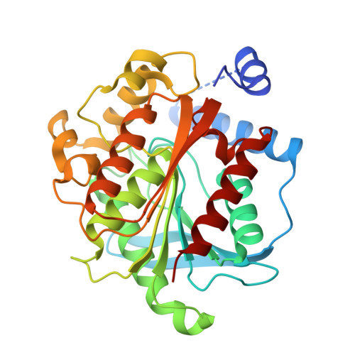
9J1V

ESTS1 phthalate ester degrading esterase from Sulfobacillus acidophilus in complex with Monomethyl phthalate


Experimental Data Snapshot

  • Method: X-RAY DIFFRACTION
  • Resolution: 2.00 Å
  • R-Value Free: 0.201 
  • R-Value Work: 0.108 

Starting Model: in silico
View more details

wwPDB Validation   3D Report Full Report


Ligand Structure Quality Assessment 


This is version 1.0 of the entry. See complete history


Literature

Mechanistic and structural insights into EstS1 esterase: A potent broad-spectrum phthalate diester degrading enzyme.

Verma, S.Choudhary, S.Amith Kumar, K.Mahto, J.K.Vamsi K, A.K.Mishra, I.Prakash, V.B.Sircar, D.Tomar, S.Kumar Sharma, A.Singla, J.Kumar, P.

(2024) Structure 

  • DOI: https://doi.org/10.1016/j.str.2024.11.006
  • Primary Citation of Related Structures:  
    8W98, 8ZE9, 9J1E, 9J1G, 9J1V, 9J57, 9J58, 9J59, 9J5A, 9J5B, 9J5C, 9J5D

  • PubMed Abstract: 

    Phthalate diesters are important pollutants and act as endocrine disruptors. While certain bacterial esterases have been identified for phthalate diesters degradation to monoesters, their structural and mechanistic characteristics remain largely unexplored. Here, we highlight the potential of the thermostable and pH-tolerant EstS1 esterase from Sulfobacillus acidophilus DSM10332 to degrade high molecular weight bis(2-ethylhexyl) phthalate (DEHP) by combining biophysical and biochemical approaches along with high-resolution EstS1 crystal structures of the apo form and with bound substrates, products, and their analogs to elucidate its mechanism. The catalytic tunnel mediates entry and exit of the substrate and product, respectively. The centralized Ser-His-Asp triad performs catalysis by a bi-bi ping-pong mechanism, forming a tetrahedral intermediate. Mutagenesis analysis showed that the Met207Ala mutation abolished DEHP binding at the active site, confirming its essential role in supporting catalysis. These findings underscore EstS1 as a promising tool for advancing technologies aimed at phthalate diesters biodegradation.


  • Organizational Affiliation

    Department of Biosciences and Bioengineering, IIT Roorkee, Roorkee, Uttarakhand 247667, India.


Macromolecules
Find similar proteins by:  (by identity cutoff)  |  3D Structure
Entity ID: 1
MoleculeChains Sequence LengthOrganismDetailsImage
Alpha/beta hydrolase fold-3 domain-containing protein310Sulfobacillus acidophilus DSM 10332Mutation(s): 0 
Gene Names: Sulac_0033
EC: 3.1.1.3
UniProt
Find proteins for G8TV28 (Sulfobacillus acidophilus (strain ATCC 700253 / DSM 10332 / NAL))
Explore G8TV28 
Go to UniProtKB:  G8TV28
Entity Groups  
Sequence Clusters30% Identity50% Identity70% Identity90% Identity95% Identity100% Identity
UniProt GroupG8TV28
Sequence Annotations
Expand
  • Reference Sequence
Small Molecules
Ligands 2 Unique
IDChains Name / Formula / InChI Key2D Diagram3D Interactions
R3O (Subject of Investigation/LOI)
Query on R3O

Download Ideal Coordinates CCD File 
B [auth A],
E [auth A],
K [auth A]
2-methoxycarbonylbenzoic acid
C9 H8 O4
FNJSWIPFHMKRAT-UHFFFAOYSA-N
EDO
Query on EDO

Download Ideal Coordinates CCD File 
C [auth A]
D [auth A]
F [auth A]
G [auth A]
H [auth A]
C [auth A],
D [auth A],
F [auth A],
G [auth A],
H [auth A],
I [auth A],
J [auth A]
1,2-ETHANEDIOL
C2 H6 O2
LYCAIKOWRPUZTN-UHFFFAOYSA-N
Experimental Data & Validation

Experimental Data

  • Method: X-RAY DIFFRACTION
  • Resolution: 2.00 Å
  • R-Value Free: 0.201 
  • R-Value Work: 0.108 
  • Space Group: P 63
Unit Cell:
Length ( Å )Angle ( ˚ )
a = 107.984α = 90
b = 107.984β = 90
c = 44.782γ = 120
Software Package:
Software NamePurpose
REFMACrefinement
CrysalisProdata reduction
CrysalisProdata scaling
MOLREPphasing

Structure Validation

View Full Validation Report


Ligand Structure Quality Assessment 


Entry History & Funding Information

Deposition Data


Funding OrganizationLocationGrant Number
Department of Science & Technology (DST, India)IndiaDST/TMD/EWO/WTI/2K19/EWFH/2019/8 (G)

Revision History  (Full details and data files)

  • Version 1.0: 2024-12-25
    Type: Initial release