


Crystal structure of Pseudopedobacter saltans GH43 beta-xylosidase in complex with xylose.
Vishwakarma, P., Sachdeva, E., Ethayathulla, A.S., Goyal, A., Singh, T.P., Kaur, P.To be published.
Experimental Data Snapshot
Starting Model: experimental
View more details
Entity ID: 1 | |||||
|---|---|---|---|---|---|
| Molecule | Chains | Sequence Length | Organism | Details | Image |
| Xylan 1,4-beta-xylosidase | 432 | Pseudopedobacter saltans DSM 12145 | Mutation(s): 0 Gene Names: Pedsa_2569 EC: 3.2.1.37 | 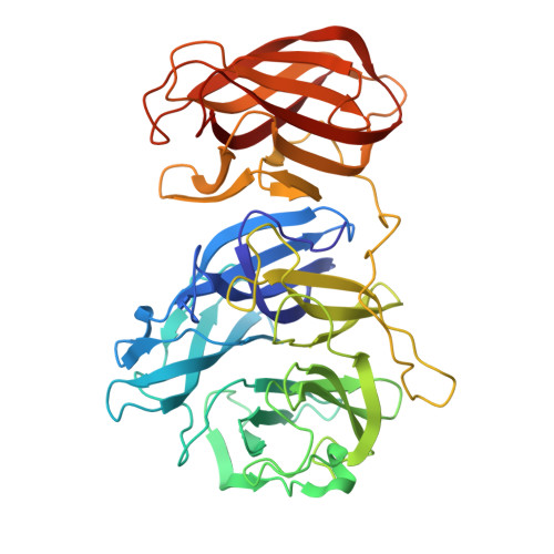 | |
UniProt | |||||
Find proteins for F0S5E9 (Pseudopedobacter saltans (strain ATCC 51119 / DSM 12145 / JCM 21818 / CCUG 39354 / LMG 10337 / NBRC 100064 / NCIMB 13643)) Explore F0S5E9 Go to UniProtKB: F0S5E9 | |||||
Entity Groups | |||||
| Sequence Clusters | 30% Identity50% Identity70% Identity90% Identity95% Identity100% Identity | ||||
| UniProt Group | F0S5E9 | ||||
Sequence AnnotationsExpand | |||||
| |||||
| Ligands 2 Unique | |||||
|---|---|---|---|---|---|
| ID | Chains | Name / Formula / InChI Key | 2D Diagram | 3D Interactions | |
| XLS (Subject of Investigation/LOI) Query on XLS | CA [auth G] FA [auth H] J [auth A] N [auth B] Q [auth C] | D-xylose C5 H10 O5 PYMYPHUHKUWMLA-VPENINKCSA-N |  | ||
| CA (Subject of Investigation/LOI) Query on CA | AA [auth G] BA [auth G] DA [auth H] EA [auth H] I [auth A] | CALCIUM ION Ca BHPQYMZQTOCNFJ-UHFFFAOYSA-N |  | ||
| Length ( Å ) | Angle ( ˚ ) |
|---|---|
| a = 73.585 | α = 102.818 |
| b = 89.435 | β = 94.019 |
| c = 138.443 | γ = 97.882 |
| Software Name | Purpose |
|---|---|
| REFMAC | refinement |
| iMOSFLM | data reduction |
| Aimless | data scaling |
| PHASER | phasing |
| Funding Organization | Location | Grant Number |
|---|---|---|
| Department of Biotechnology (DBT, India) | India | -- |


