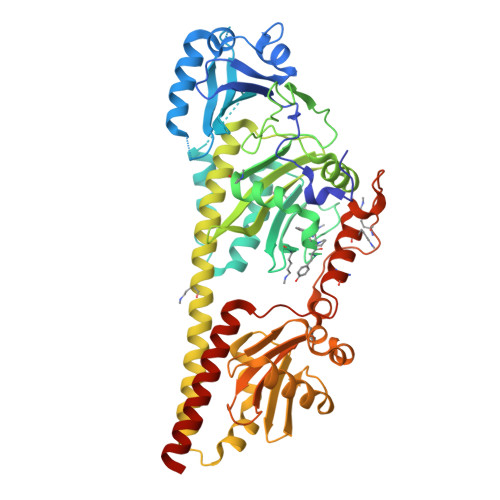
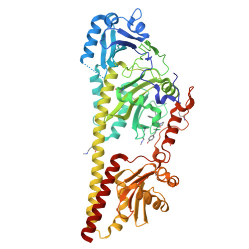
9G8D

Crystal structure of the photosensory core module (PCM) of a cyano-phenylalanine mutant oCNF192 of the bathy phytochrome Agp2 from Agrobacterium fabrum in the Pfr state.


Experimental Data Snapshot

  • Method: X-RAY DIFFRACTION
  • Resolution: 2.06 Å
  • R-Value Free: 0.224 
  • R-Value Work: 0.186 
  • R-Value Observed: 0.188 

Starting Model: experimental
View more details

wwPDB Validation   3D Report Full Report


Ligand Structure Quality Assessment 


This is version 1.1 of the entry. See complete history


Literature

Hydrogen Bonding and Noncovalent Electric Field Effects in the Photoconversion of a Phytochrome.

Nguyen, A.D.Michael, N.Sauthof, L.von Sass, J.Hoang, O.T.Schmidt, A.La Greca, M.Schlesinger, R.Budisa, N.Scheerer, P.Mroginski, M.A.Kraskov, A.Hildebrandt, P.

(2024) J Phys Chem B 128: 11644-11657

  • DOI: https://doi.org/10.1021/acs.jpcb.4c06419
  • Primary Citation of Related Structures:  
    9G8C, 9G8D

  • PubMed Abstract: 

    A profound understanding of protein structure and mechanism requires dedicated experimental and theoretical tools to elucidate electrostatic and hydrogen bonding interactions in proteins. In this work, we employed an approach to disentangle noncovalent and hydrogen-bonding electric field changes during the reaction cascade of a multidomain protein, i.e., the phytochrome Agp2. The approach exploits the spectroscopic properties of nitrile probes commonly used as reporter groups of the vibrational Stark effect. These probes were introduced into the protein through site-specific incorporation of noncanonical amino acids resulting in four variants with different positions and orientations of the nitrile groups. All substitutions left structures and the reaction mechanism unchanged. Structural models of the dark states (Pfr) were used to evaluate the total electric field at the nitrile label and its transition dipole moment. These quantities served as an internal standard to calculate the respective properties of the photoinduced products (Lumi-F, Meta-F, and Pr) based on the relative intensities of the nitrile stretching bands. In most cases, the spectral analysis revealed two substates with a nitrile in a hydrogen-bonded or hydrophobic environment. Using frequencies and intensities, we managed to extract the noncovalent contribution of the electric field from the individual substates. This analysis resulted in profiles of the noncovalent and hydrogen-bond-related electric fields during the photoinduced reaction cascade of Agp2. These profiles, which vary significantly among the four variants due to the different positions and orientations of the nitrile probes, were discussed in the context of the molecular events along the Pfr → Pr reaction cascade.


  • Organizational Affiliation

    Institut für Chemie, Sekr. C7, Technische Universität Berlin, Straße des 17. Juni 115, Berlin D-10623, Germany.


Macromolecules
Find similar proteins by:  (by identity cutoff)  |  3D Structure
Entity ID: 1
MoleculeChains Sequence LengthOrganismDetailsImage
histidine kinase507Agrobacterium fabrum str. C58Mutation(s): 0 
Gene Names: Atu2165
EC: 2.7.13.3
UniProt
Find proteins for A9CI81 (Agrobacterium fabrum (strain C58 / ATCC 33970))
Explore A9CI81 
Go to UniProtKB:  A9CI81
Entity Groups  
Sequence Clusters30% Identity50% Identity70% Identity90% Identity95% Identity100% Identity
UniProt GroupA9CI81
Sequence Annotations
Expand
  • Reference Sequence
Find similar proteins by:  (by identity cutoff)  |  3D Structure
Entity ID: 2
MoleculeChains Sequence LengthOrganismDetailsImage
histidine kinase507Agrobacterium fabrum str. C58Mutation(s): 0 
Gene Names: Atu2165
EC: 2.7.13.3
UniProt
Find proteins for A9CI81 (Agrobacterium fabrum (strain C58 / ATCC 33970))
Explore A9CI81 
Go to UniProtKB:  A9CI81
Entity Groups  
Sequence Clusters30% Identity50% Identity70% Identity90% Identity95% Identity100% Identity
UniProt GroupA9CI81
Sequence Annotations
Expand
  • Reference Sequence
Small Molecules
Ligands 4 Unique
IDChains Name / Formula / InChI Key2D Diagram3D Interactions
EL5 (Subject of Investigation/LOI)
Query on EL5

Download Ideal Coordinates CCD File 
E [auth A],
J [auth B]
3-[(2Z)-2-({3-(2-carboxyethyl)-5-[(E)-(4-ethenyl-3-methyl-5-oxo-1,5-dihydro-2H-pyrrol-2-ylidene)methyl]-4-methyl-1H-pyrrol-2-yl}methylidene)-5-{(Z)-[(3E,4S)-3-ethylidene-4-methyl-5-oxopyrrolidin-2-ylidene]methyl}-4-methyl-2H-pyrrol-3-yl]propanoic acid
C33 H36 N4 O6
SNHIGJASYQUMKZ-IDFYGOSVSA-N
MPD
Query on MPD

Download Ideal Coordinates CCD File 
D [auth A],
I [auth B]
(4S)-2-METHYL-2,4-PENTANEDIOL
C6 H14 O2
SVTBMSDMJJWYQN-YFKPBYRVSA-N
PEG
Query on PEG

Download Ideal Coordinates CCD File 
C [auth A],
H [auth B]
DI(HYDROXYETHYL)ETHER
C4 H10 O3
MTHSVFCYNBDYFN-UHFFFAOYSA-N
CL
Query on CL

Download Ideal Coordinates CCD File 
F [auth A],
G [auth A]
CHLORIDE ION
Cl
VEXZGXHMUGYJMC-UHFFFAOYSA-M
Modified Residues  1 Unique
IDChains TypeFormula2D DiagramParent
MLZ
Query on MLZ
A
L-PEPTIDE LINKINGC7 H16 N2 O2LYS
Experimental Data & Validation

Experimental Data

  • Method: X-RAY DIFFRACTION
  • Resolution: 2.06 Å
  • R-Value Free: 0.224 
  • R-Value Work: 0.186 
  • R-Value Observed: 0.188 
  • Space Group: P 21 21 21
Unit Cell:
Length ( Å )Angle ( ˚ )
a = 74.644α = 90
b = 93.577β = 90
c = 173.945γ = 90
Software Package:
Software NamePurpose
REFMACrefinement
XDSdata reduction
Aimlessdata scaling
PHASERphasing

Structure Validation

View Full Validation Report


Ligand Structure Quality Assessment 


Entry History & Funding Information

Deposition Data


Funding OrganizationLocationGrant Number
German Research Foundation (DFG)GermanyCRC1078 - project number 221545957 - subproject B06
German Research Foundation (DFG)GermanyEXC 311 2008/1 - UniSysCat - 390540038 - Research Unit E

Revision History  (Full details and data files)

  • Version 1.0: 2024-11-27
    Type: Initial release
  • Version 1.1: 2024-12-11
    Changes: Database references