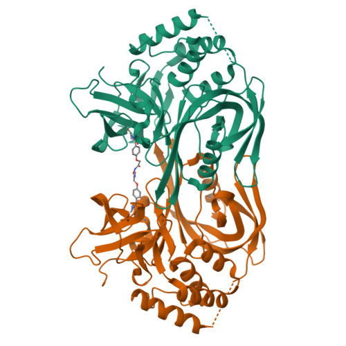
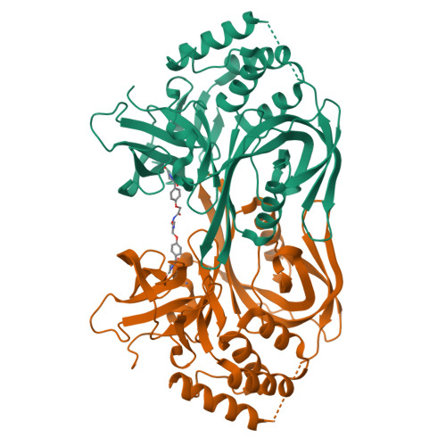
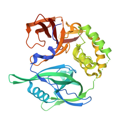
RCSB PDB - 9GAO: Crystal structure of CRBNmidi in complex with 2-(4-(2,6-dioxopiperidin-3-yl)phenoxy)-N-methylacetamide

 9GAO

Crystal structure of CRBNmidi in complex with 2-(4-(2,6-dioxopiperidin-3-yl)phenoxy)-N-methylacetamide


Experimental Data Snapshot

  • Method: X-RAY DIFFRACTION
  • Resolution: 1.95 Å
  • R-Value Free: 0.286 
  • R-Value Work: 0.252 
  • R-Value Observed: 0.254 

Starting Model: experimental
View more details

wwPDB Validation   3D Report Full Report


Ligand Structure Quality Assessment 

Created with Raphaël 2.3.0Worse 01 BetterLigand structure goodness of fit to experimental dataBest fitted A1IJMClick on this verticalbar to view details

This is version 1.1 of the entry. See complete history


Literature

Design of a Cereblon construct for crystallographic and biophysical studies of protein degraders.

Kroupova, A.Spiteri, V.A.Rutter, Z.J.Furihata, H.Darren, D.Ramachandran, S.Chakraborti, S.Haubrich, K.Pethe, J.Gonzales, D.Wijaya, A.J.Rodriguez-Rios, M.Sturbaut, M.Lynch, D.M.Farnaby, W.Nakasone, M.A.Zollman, D.Ciulli, A.

(2024) Nat Commun 15: 8885-8885

  • DOI: https://doi.org/10.1038/s41467-024-52871-9
  • Primary Citation of Related Structures:  
    8RQ1, 8RQ8, 8RQ9, 8RQA, 8RQC, 9GAO

  • PubMed Abstract: 

    The ubiquitin E3 ligase cereblon (CRBN) is the target of therapeutic drugs thalidomide and lenalidomide and is recruited by most targeted protein degraders (PROTACs and molecular glues) in clinical development. Biophysical and structural investigation of CRBN has been limited by current constructs that either require co-expression with the adaptor DDB1 or inadequately represent full-length protein, with high-resolution structures of degrader ternary complexes remaining rare. We present the design of CRBN midi , a construct that readily expresses from E. coli with high yields as soluble, stable protein without DDB1. We benchmark CRBN midi for wild-type functionality through a suite of biophysical techniques and solve high-resolution co-crystal structures of its binary and ternary complexes with degraders. We qualify CRBN midi as an enabling tool to accelerate structure-based discovery of the next generation of CRBN based therapeutics.


  • Organizational Affiliation

    Centre for Targeted Protein Degradation, School of Life Sciences, University of Dundee, Dundee, DD1 5JJ, UK.


Macromolecules
Find similar proteins by:  (by identity cutoff)  |  3D Structure
Entity ID: 1
MoleculeChains Sequence LengthOrganismDetailsImage
Protein cereblonA,
B [auth C]
329Homo sapiensMutation(s): 12 
Gene Names: CRBNAD-006
UniProt & NIH Common Fund Data Resources
Find proteins for Q96SW2 (Homo sapiens)
Explore Q96SW2 
Go to UniProtKB:  Q96SW2
PHAROS:  Q96SW2
GTEx:  ENSG00000113851 
Entity Groups  
Sequence Clusters30% Identity50% Identity70% Identity90% Identity95% Identity100% Identity
UniProt GroupQ96SW2
Sequence Annotations
Expand
  • Reference Sequence
Experimental Data & Validation

Experimental Data

  • Method: X-RAY DIFFRACTION
  • Resolution: 1.95 Å
  • R-Value Free: 0.286 
  • R-Value Work: 0.252 
  • R-Value Observed: 0.254 
  • Space Group: P 21 21 21
Unit Cell:
Length ( Å )Angle ( ˚ )
a = 53.507α = 90
b = 94.862β = 90
c = 147.952γ = 90
Software Package:
Software NamePurpose
PHENIXrefinement
autoPROCdata reduction
autoPROCdata scaling
PHASERphasing
Cootmodel building

Structure Validation

View Full Validation Report


Ligand Structure Quality Assessment 

Created with Raphaël 2.3.0Worse 01 BetterLigand structure goodness of fit to experimental dataBest fitted A1IJMClick on this verticalbar to view details

Entry History & Funding Information

Deposition Data


Funding OrganizationLocationGrant Number
Other private--

Revision History  (Full details and data files)

  • Version 1.0: 2024-10-02
    Type: Initial release
  • Version 1.1: 2024-10-30
    Changes: Advisory, Database references, Structure summary