1OC5

D405N mutant of the CELLOBIOHYDROLASE CEL6A FROM HUMICOLA INSOLENS in complex with methyl-cellobiosyl-4-deoxy-4-thio-beta-D-cellobioside


Experimental Data Snapshot

  • Method: X-RAY DIFFRACTION
  • Resolution: 1.70 Å
  • R-Value Free: 0.168 
  • R-Value Work: 0.127 
  • R-Value Observed: 0.129 

wwPDB Validation   3D Report Full Report


Ligand Structure Quality Assessment 


This is version 2.1 of the entry. See complete history


Literature

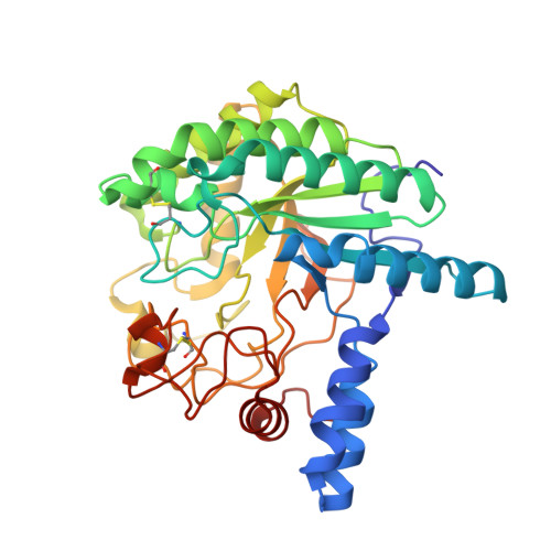
Structural Basis for Ligand Binding and Processivity in Cellobiohydrolase Cel6A from Humicola Insolens

Varrot, A.Frandsen, T.P.Von Ossowski, I.Boyer, V.Driguez, H.Schulein, M.Davies, G.J.

(2003) Structure 11: 855

  • DOI: https://doi.org/10.1016/s0969-2126(03)00124-2
  • Primary Citation of Related Structures:  
    1OC5, 1OC6, 1OC7, 1OCB, 1OCJ

  • PubMed Abstract: 

    The enzymatic digestion of cellulose entails intimate involvement of cellobiohydrolases, whose characteristic active-center tunnel contributes to a processive degradation of the polysaccharide. The cellobiohydrolase Cel6A displays an active site within a tunnel formed by two extended loops, which are known to open and close in response to ligand binding. Here we present five structures of wild-type and mutant forms of Cel6A from Humicola insolens in complex with nonhydrolyzable thio-oligosaccharides, at resolutions from 1.7-1.1 A, dissecting the structural accommodation of a processing substrate chain through the active center during hydrolysis. Movement of ligand is facilitated by extensive solvent-mediated interactions and through flexibility in the hydrophobic surfaces provided by a sheath of tryptophan residues.


  • Organizational Affiliation

    Department of Chemistry, University of York, Heslington, Y010 5YW, York, United Kingdom.


Macromolecules
Find similar proteins by:  (by identity cutoff)  |  3D Structure
Entity ID: 1
MoleculeChains Sequence LengthOrganismDetailsImage
CELLOBIOHYDROLASE II364Humicola insolensMutation(s): 0 
EC: 3.2.1.91
UniProt
Find proteins for Q9C1S9 (Humicola insolens)
Explore Q9C1S9 
Go to UniProtKB:  Q9C1S9
Entity Groups  
Sequence Clusters30% Identity50% Identity70% Identity90% Identity95% Identity100% Identity
UniProt GroupQ9C1S9
Sequence Annotations
Expand
  • Reference Sequence
Oligosaccharides

Help

Entity ID: 2
MoleculeChains Length2D Diagram Glycosylation3D Interactions
beta-D-glucopyranose-(1-4)-beta-D-glucopyranose-(1-4)-4-thio-beta-D-glucopyranose-(1-4)-methyl beta-D-glucopyranoside
B
4N/AN/A
Glycosylation Resources
GlyTouCan:  G15263TL
GlyCosmos:  G15263TL
Experimental Data & Validation

Experimental Data

  • Method: X-RAY DIFFRACTION
  • Resolution: 1.70 Å
  • R-Value Free: 0.168 
  • R-Value Work: 0.127 
  • R-Value Observed: 0.129 
  • Space Group: P 21 21 21
Unit Cell:
Length ( Å )Angle ( ˚ )
a = 57.504α = 90
b = 60.148β = 90
c = 97.207γ = 90
Software Package:
Software NamePurpose
REFMACrefinement
DENZOdata reduction
SCALEPACKdata scaling
AMoREphasing

Structure Validation

View Full Validation Report


Ligand Structure Quality Assessment 


Entry History 

Deposition Data

Revision History  (Full details and data files)

  • Version 1.0: 2003-07-10
    Type: Initial release
  • Version 1.1: 2011-05-08
    Changes: Version format compliance
  • Version 1.2: 2011-07-13
    Changes: Version format compliance
  • Version 2.0: 2020-07-29
    Type: Remediation
    Reason: Carbohydrate remediation
    Changes: Advisory, Atomic model, Data collection, Derived calculations, Other, Structure summary
  • Version 2.1: 2023-12-13
    Changes: Data collection, Database references, Refinement description, Structure summary