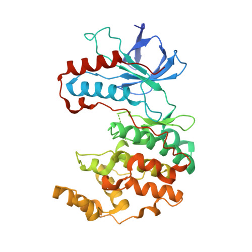
3P7A

p38 inhibitor-bound


Experimental Data Snapshot

  • Method: X-RAY DIFFRACTION
  • Resolution: 2.31 Å
  • R-Value Free: 0.298 
  • R-Value Work: 0.224 
  • R-Value Observed: 0.230 

wwPDB Validation   3D Report Full Report


Ligand Structure Quality Assessment 


This is version 1.5 of the entry. See complete history


Literature

Discovery of a novel class of non-ATP site DFG-out state p38 inhibitors utilizing computationally assisted virtual fragment-based drug design (vFBDD).

Moffett, K.Konteatis, Z.Nguyen, D.Shetty, R.Ludington, J.Fujimoto, T.Lee, K.J.Chai, X.Namboodiri, H.Karpusas, M.Dorsey, B.Guarnieri, F.Bukhtiyarova, M.Springman, E.Michelotti, E.

(2011) Bioorg Med Chem Lett 21: 7155-7165

  • DOI: https://doi.org/10.1016/j.bmcl.2011.09.078
  • Primary Citation of Related Structures:  
    3P5K, 3P78, 3P79, 3P7A, 3P7B, 3P7C

  • PubMed Abstract: 

    Discovery of a new class of DFG-out p38α kinase inhibitors with no hinge interaction is described. A computationally assisted, virtual fragment-based drug design (vFBDD) platform was utilized to identify novel non-aromatic fragments which make productive hydrogen bond interactions with Arg 70 on the αC-helix. Molecules incorporating these fragments were found to be potent inhibitors of p38 kinase. X-ray co-crystal structures confirmed the predicted binding modes. A lead compound was identified as a potent (p38α IC(50)=22 nM) and highly selective (≥ 150-fold against 150 kinase panel) DFG-out p38 kinase inhibitor.


  • Organizational Affiliation

    Ansaris, Four Valley Square, 512 East Township Line Road, Blue Bell, PA 19422, USA. kmistry@earthlink.net


Macromolecules
Find similar proteins by:  (by identity cutoff)  |  3D Structure
Entity ID: 1
MoleculeChains Sequence LengthOrganismDetailsImage
Mitogen-activated protein kinase 14366Mus musculusMutation(s): 0 
Gene Names: Mapk14Crk1Csbp1Csbp2
EC: 2.7.11.24
UniProt
Find proteins for P47811 (Mus musculus)
Explore P47811 
Go to UniProtKB:  P47811
Entity Groups  
Sequence Clusters30% Identity50% Identity70% Identity90% Identity95% Identity100% Identity
UniProt GroupP47811
Sequence Annotations
Expand
  • Reference Sequence
Small Molecules
Ligands 2 Unique
IDChains Name / Formula / InChI Key2D Diagram3D Interactions
P7A
Query on P7A

Download Ideal Coordinates CCD File 
C [auth A]1-[5-tert-butyl-2-(1,1-dioxidothiomorpholin-4-yl)thiophen-3-yl]-3-naphthalen-1-ylurea
C23 H27 N3 O3 S2
VRPGNWGNMYKKMF-UHFFFAOYSA-N
BOG
Query on BOG

Download Ideal Coordinates CCD File 
B [auth A]octyl beta-D-glucopyranoside
C14 H28 O6
HEGSGKPQLMEBJL-RKQHYHRCSA-N
Binding Affinity Annotations 
IDSourceBinding Affinity
P7A PDBBind:  3P7A IC50: 520 (nM) from 1 assay(s)
BindingDB:  3P7A IC50: 520 (nM) from 1 assay(s)
Experimental Data & Validation

Experimental Data

  • Method: X-RAY DIFFRACTION
  • Resolution: 2.31 Å
  • R-Value Free: 0.298 
  • R-Value Work: 0.224 
  • R-Value Observed: 0.230 
  • Space Group: P 21 21 21
Unit Cell:
Length ( Å )Angle ( ˚ )
a = 65.14α = 90
b = 74.51β = 90
c = 74.41γ = 90
Software Package:
Software NamePurpose
REFMACrefinement
CNSrefinement
HKL-2000data reduction
HKL-2000data scaling
CNSphasing
HKL-2000data collection

Structure Validation

View Full Validation Report


Ligand Structure Quality Assessment 


Entry History 

Deposition Data

Revision History  (Full details and data files)

  • Version 1.0: 2011-10-12
    Type: Initial release
  • Version 1.1: 2011-11-09
    Changes: Database references
  • Version 1.2: 2012-01-25
    Changes: Database references
  • Version 1.3: 2019-07-17
    Changes: Data collection, Refinement description
  • Version 1.4: 2020-07-29
    Type: Remediation
    Reason: Carbohydrate remediation
    Changes: Data collection, Database references, Derived calculations, Structure summary
  • Version 1.5: 2024-02-21
    Changes: Data collection, Database references, Structure summary