3VED | pdb_00003ved

Rhodococcus jostii RHA1 DypB D153H variant in complex with heme


Experimental Data Snapshot

  • Method: X-RAY DIFFRACTION
  • Resolution: 2.50 Å
  • R-Value Free: 
    0.218 (Depositor), 0.210 (DCC) 
  • R-Value Work: 
    0.184 (Depositor), 0.180 (DCC) 
  • R-Value Observed: 
    0.185 (Depositor) 

Starting Model: experimental
View more details

wwPDB Validation   3D Report Full Report


Ligand Structure Quality Assessment 

Created with Raphaël 2.3.0Worse 01 BetterLigand structure goodness of fit to experimental dataBest fitted HEMClick on this verticalbar to view details

This is version 1.3 of the entry. See complete history


Literature

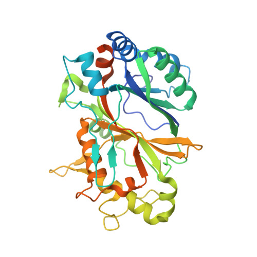
Distal heme pocket residues of B-type dye-decolorizing peroxidase: arginine but not aspartate is essential for peroxidase activity.

Singh, R.Grigg, J.C.Armstrong, Z.Murphy, M.E.Eltis, L.D.

(2012) J Biological Chem 287: 10623-10630

  • DOI: https://doi.org/10.1074/jbc.M111.332171
  • Primary Citation of Related Structures:  
    3VEC, 3VED, 3VEE, 3VEF, 3VEG

  • PubMed Abstract: 

    DypB from Rhodococcus jostii RHA1 is a bacterial dye-decolorizing peroxidase (DyP) that oxidizes lignin and Mn(II). Three residues interact with the iron-bound solvent species in ferric DypB: Asn-246 and the conserved Asp-153 and Arg-244. Substitution of either Asp-153 or Asn-246 with alanine minimally affected the second order rate constant for Compound I formation (k(1) ∼ 10(5) M(-1)s(-1)) and the specificity constant (k(cat)/K(m)) for H(2)O(2). Even in the D153A/N246A double variant, these values were reduced less than 30-fold. However, these substitutions dramatically reduced the stability of Compound I (t(1/2) ∼ 0.13 s) as compared with the wild-type enzyme (540 s). By contrast, substitution of Arg-244 with leucine abolished the peroxidase activity, and heme iron of the variant showed a pH-dependent transition from high spin (pH 5) to low spin (pH 8.5). Two variants were designed to mimic the plant peroxidase active site: D153H, which was more than an order of magnitude less reactive with H(2)O(2), and N246H, which had no detectable peroxidase activity. X-ray crystallographic studies revealed that structural changes in the variants are confined to the distal heme environment. The data establish an essential role for Arg-244 in Compound I formation in DypB, possibly through charge stabilization and proton transfer. The principle roles of Asp-153 and Asn-246 appear to be in modulating the subsequent reactivity of Compound I. These results expand the range of residues known to catalyze Compound I formation in heme peroxidases.


  • Organizational Affiliation

    Department of Microbiology and Immunology, Life Sciences Institute, University of British Columbia, Vancouver, British Columbia V6T 1Z3, Canada.


Macromolecules
Find similar proteins by:  (by identity cutoff)  |  3D Structure
Entity ID: 1
MoleculeChains Sequence LengthOrganismDetailsImage
DypB
A, B, C
353Rhodococcus jostii RHA1Mutation(s): 1 
Gene Names: dypBRHA1_ro02407
EC: 1.11.1
UniProt
Find proteins for Q0SE24 (Rhodococcus jostii (strain RHA1))
Explore Q0SE24 
Go to UniProtKB:  Q0SE24
Entity Groups  
Sequence Clusters30% Identity50% Identity70% Identity90% Identity95% Identity100% Identity
UniProt GroupQ0SE24
Sequence Annotations
Expand
  • Reference Sequence
Small Molecules
Ligands 3 Unique
IDChains Name / Formula / InChI Key2D Diagram3D Interactions
HEM
Query on HEM

Download Ideal Coordinates CCD File 
D [auth A],
H [auth B],
K [auth C]
PROTOPORPHYRIN IX CONTAINING FE
C34 H32 Fe N4 O4
KABFMIBPWCXCRK-RGGAHWMASA-L
GOL
Query on GOL

Download Ideal Coordinates CCD File 
F [auth A]GLYCEROL
C3 H8 O3
PEDCQBHIVMGVHV-UHFFFAOYSA-N
CL
Query on CL

Download Ideal Coordinates CCD File 
E [auth A]
G [auth A]
I [auth B]
J [auth B]
L [auth C]
CHLORIDE ION
Cl
VEXZGXHMUGYJMC-UHFFFAOYSA-M
Experimental Data & Validation

Experimental Data

  • Method: X-RAY DIFFRACTION
  • Resolution: 2.50 Å
  • R-Value Free:  0.218 (Depositor), 0.210 (DCC) 
  • R-Value Work:  0.184 (Depositor), 0.180 (DCC) 
  • R-Value Observed: 0.185 (Depositor) 
Space Group: P 32 2 1
Unit Cell:
Length ( Å )Angle ( ˚ )
a = 132.642α = 90
b = 132.642β = 90
c = 159.61γ = 120
Software Package:
Software NamePurpose
SCALEPACKdata scaling
REFMACrefinement
PDB_EXTRACTdata extraction
MxDCdata collection
HKL-2000data reduction
HKL-2000data scaling
REFMACphasing

Structure Validation

View Full Validation Report


Ligand Structure Quality Assessment 

Created with Raphaël 2.3.0Worse 01 BetterLigand structure goodness of fit to experimental dataBest fitted HEMClick on this verticalbar to view details

Entry History 

Deposition Data

Revision History  (Full details and data files)

  • Version 1.0: 2012-01-18
    Type: Initial release
  • Version 1.1: 2012-08-29
    Changes: Database references
  • Version 1.2: 2017-11-08
    Changes: Refinement description
  • Version 1.3: 2023-09-13
    Changes: Data collection, Database references, Derived calculations, Refinement description