5V7N

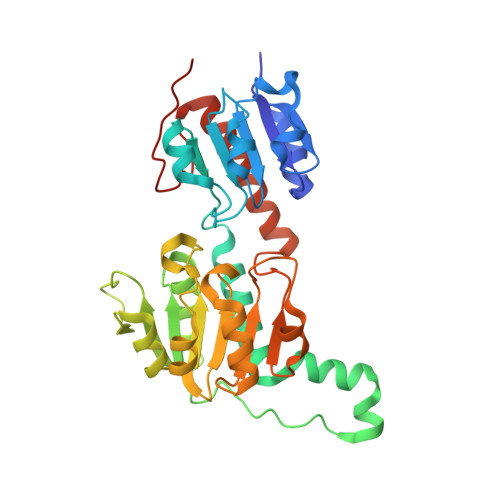
Crystal structure of NADPH-dependent glyoxylate/hydroxypyruvate reductase SMc04462 (SmGhrB) from Sinorhizobium meliloti in complex with NADP and 2-Keto-D-gluconic acid


Experimental Data Snapshot

  • Method: X-RAY DIFFRACTION
  • Resolution: 1.75 Å
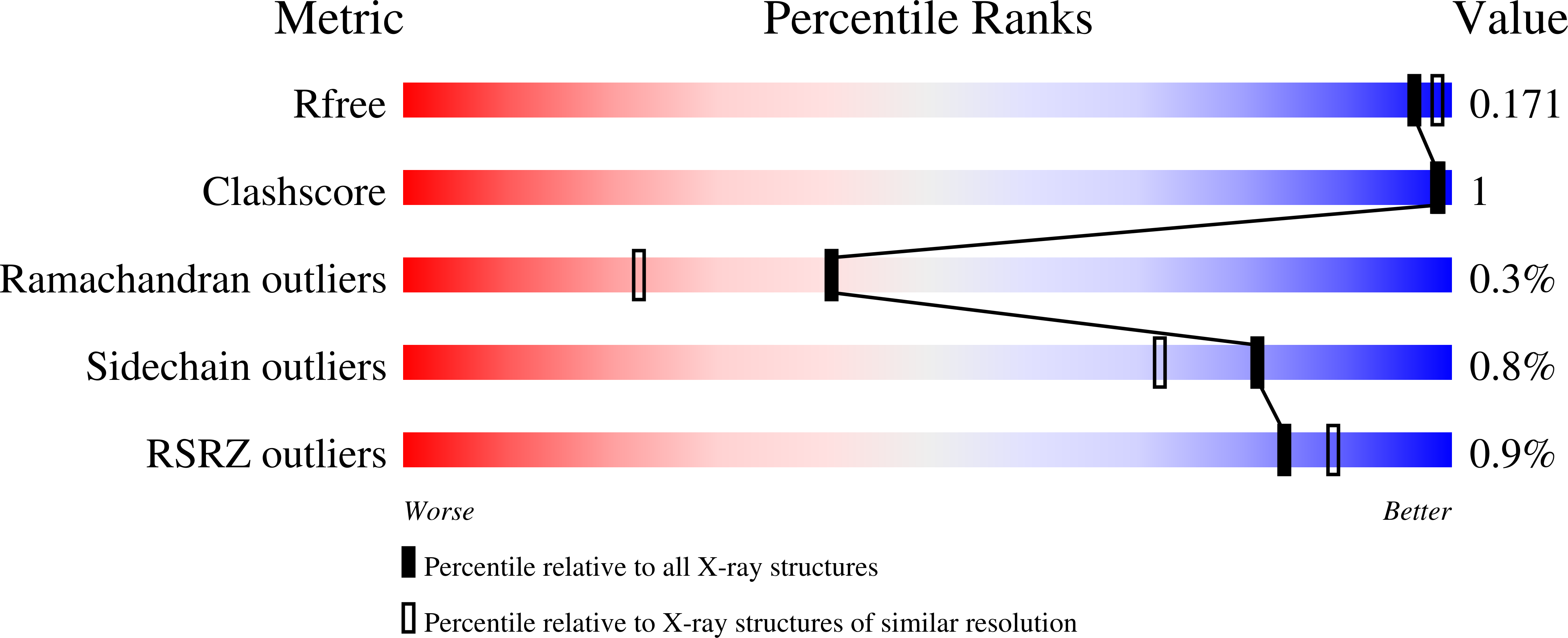
  • R-Value Free: 0.167 
  • R-Value Work: 0.144 
  • R-Value Observed: 0.145 

Starting Model: experimental
View more details

wwPDB Validation   3D Report Full Report


Ligand Structure Quality Assessment 


This is version 2.1 of the entry. See complete history


Literature

Structural, Biochemical, and Evolutionary Characterizations of Glyoxylate/Hydroxypyruvate Reductases Show Their Division into Two Distinct Subfamilies.

Kutner, J.Shabalin, I.G.Matelska, D.Handing, K.B.Gasiorowska, O.Sroka, P.Gorna, M.W.Ginalski, K.Wozniak, K.Minor, W.

(2018) Biochemistry 57: 963-977

  • DOI: https://doi.org/10.1021/acs.biochem.7b01137
  • Primary Citation of Related Structures:  
    4WEQ, 4Z0P, 5J23, 5UNN, 5UOG, 5V6Q, 5V72, 5V7G, 5V7N

  • PubMed Abstract: 

    The d-2-hydroxyacid dehydrogenase (2HADH) family illustrates a complex evolutionary history with multiple lateral gene transfers and gene duplications and losses. As a result, the exact functional annotation of individual members can be extrapolated to a very limited extent. Here, we revise the previous simplified view on the classification of the 2HADH family; specifically, we show that the previously delineated glyoxylate/hydroxypyruvate reductase (GHPR) subfamily consists of two evolutionary separated GHRA and GHRB subfamilies. We compare two representatives of these subfamilies from Sinorhizobium meliloti (SmGhrA and SmGhrB), employing a combination of biochemical, structural, and bioinformatics approaches. Our kinetic results show that both enzymes reduce several 2-ketocarboxylic acids with overlapping, but not equivalent, substrate preferences. SmGhrA and SmGhrB show highest activity with glyoxylate and hydroxypyruvate, respectively; in addition, only SmGhrB reduces 2-keto-d-gluconate, and only SmGhrA reduces pyruvate (with low efficiency). We present nine crystal structures of both enzymes in apo forms and in complexes with cofactors and substrates/substrate analogues. In particular, we determined a crystal structure of SmGhrB with 2-keto-d-gluconate, which is the biggest substrate cocrystallized with a 2HADH member. The structures reveal significant differences between SmGhrA and SmGhrB, both in the overall structure and within the substrate-binding pocket, offering insight into the molecular basis for the observed substrate preferences and subfamily differences. In addition, we provide an overview of all GHRA and GHRB structures complexed with a ligand in the active site.


  • Organizational Affiliation

    Department of Molecular Physiology and Biological Physics, University of Virginia , 1340 Jefferson Park Avenue, Charlottesville, Virginia 22908, United States.


Macromolecules
Find similar proteins by:  (by identity cutoff)  |  3D Structure
Entity ID: 1
MoleculeChains Sequence LengthOrganismDetailsImage
NADPH-dependent glyoxylate/hydroxypyruvate reductase
A, B, C, D
323Sinorhizobium meliloti 1021Mutation(s): 0 
Gene Names: SMc04462
EC: 1.1.1.81
UniProt
Find proteins for Q92LZ4 (Rhizobium meliloti (strain 1021))
Explore Q92LZ4 
Go to UniProtKB:  Q92LZ4
Entity Groups  
Sequence Clusters30% Identity50% Identity70% Identity90% Identity95% Identity100% Identity
UniProt GroupQ92LZ4
Sequence Annotations
Expand
  • Reference Sequence
Small Molecules
Ligands 8 Unique
IDChains Name / Formula / InChI Key2D Diagram3D Interactions
NAP
Query on NAP

Download Ideal Coordinates CCD File 
AA [auth D],
E [auth A],
N [auth B],
V [auth C]
NADP NICOTINAMIDE-ADENINE-DINUCLEOTIDE PHOSPHATE
C21 H28 N7 O17 P3
XJLXINKUBYWONI-NNYOXOHSSA-N
PG4
Query on PG4

Download Ideal Coordinates CCD File 
K [auth A]TETRAETHYLENE GLYCOL
C8 H18 O5
UWHCKJMYHZGTIT-UHFFFAOYSA-N
8YV
Query on 8YV

Download Ideal Coordinates CCD File 
BA [auth D],
F [auth A],
O [auth B],
W [auth C]
2-keto-D-gluconic acid
C6 H10 O7
VBUYCZFBVCCYFD-JJYYJPOSSA-N
PEG
Query on PEG

Download Ideal Coordinates CCD File 
DA [auth D],
H [auth A]
DI(HYDROXYETHYL)ETHER
C4 H10 O3
MTHSVFCYNBDYFN-UHFFFAOYSA-N
GOL
Query on GOL

Download Ideal Coordinates CCD File 
GA [auth D],
S [auth B]
GLYCEROL
C3 H8 O3
PEDCQBHIVMGVHV-UHFFFAOYSA-N
CL
Query on CL

Download Ideal Coordinates CCD File 
EA [auth D]
FA [auth D]
I [auth A]
J [auth A]
Q [auth B]
EA [auth D],
FA [auth D],
I [auth A],
J [auth A],
Q [auth B],
R [auth B],
Y [auth C]
CHLORIDE ION
Cl
VEXZGXHMUGYJMC-UHFFFAOYSA-M
MG
Query on MG

Download Ideal Coordinates CCD File 
HA [auth D]
IA [auth D]
L [auth A]
M [auth A]
T [auth B]
HA [auth D],
IA [auth D],
L [auth A],
M [auth A],
T [auth B],
U [auth B],
Z [auth C]
MAGNESIUM ION
Mg
JLVVSXFLKOJNIY-UHFFFAOYSA-N
NA
Query on NA

Download Ideal Coordinates CCD File 
CA [auth D],
G [auth A],
P [auth B],
X [auth C]
SODIUM ION
Na
FKNQFGJONOIPTF-UHFFFAOYSA-N
Experimental Data & Validation

Experimental Data

  • Method: X-RAY DIFFRACTION
  • Resolution: 1.75 Å
  • R-Value Free: 0.167 
  • R-Value Work: 0.144 
  • R-Value Observed: 0.145 
  • Space Group: H 3
  • Diffraction Data: https://doi.org/10.18430/M35V7N
Unit Cell:
Length ( Å )Angle ( ˚ )
a = 175.902α = 90
b = 175.902β = 90
c = 135.336γ = 120
Software Package:
Software NamePurpose
MD2data collection
HKL-3000data reduction
HKL-3000data scaling
MOLREPphasing
REFMACrefinement
PDB_EXTRACTdata extraction
DENZOdata reduction
SCALEPACKdata scaling

Structure Validation

View Full Validation Report


Ligand Structure Quality Assessment 


Entry History & Funding Information

Deposition Data


Funding OrganizationLocationGrant Number
National Institutes of Health/National Institute of General Medical Sciences (NIH/NIGMS)United StatesGM094662
National Institutes of Health/National Institute of General Medical Sciences (NIH/NIGMS)United StatesGM117080
National Institutes of Health/National Human Genome Research Institute (NIH/NHGRI)United StatesHG008424

Revision History  (Full details and data files)

  • Version 1.0: 2017-03-29
    Type: Initial release
  • Version 1.1: 2017-09-13
    Changes: Author supporting evidence, Refinement description
  • Version 1.2: 2018-05-16
    Changes: Data collection, Database references, Structure summary
  • Version 1.3: 2019-11-27
    Changes: Author supporting evidence
  • Version 2.0: 2020-07-01
    Changes: Atomic model, Structure summary
  • Version 2.1: 2023-10-04
    Changes: Data collection, Database references, Derived calculations, Refinement description