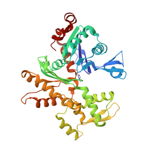
3HBT

The structure of native G-actin


Experimental Data Snapshot

  • Method: X-RAY DIFFRACTION
  • Resolution: 2.70 Å
  • R-Value Free: 0.257 
  • R-Value Work: 0.202 
  • R-Value Observed: 0.205 

Starting Model: experimental
View more details

wwPDB Validation   3D Report Full Report


Ligand Structure Quality Assessment 


This is version 1.2 of the entry. See complete history


Literature

The structure of native G-actin

Wang, H.Robinson, R.C.Burtnick, L.D.

(2010) Cytoskeleton (Hoboken) 67: 456-465

  • DOI: https://doi.org/10.1002/cm.20458
  • Primary Citation of Related Structures:  
    3HBT

  • PubMed Abstract: 

    Heat shock proteins act as cytoplasmic chaperones to ensure correct protein folding and prevent protein aggregation. The presence of stoichiometric amounts of one such heat shock protein, Hsp27, in supersaturated solutions of unmodified G-actin leads to crystallization, in preference to polymerization, of the actin. Hsp27 is not evident in the resulting crystal structure. Thus, for the first time, we present the structure of G-actin in a form that is devoid of polymerization-deterring chemical modifications or binding partners, either of which may alter its conformation. The structure contains a calcium ion and ATP within a closed nucleotide-binding cleft, and the D-loop is disordered. This native G-actin structure invites comparison with the current F-actin model in order to understand the structural implications for actin polymerization. In particular, this analysis suggests a mechanism by which the bound cation coordinates conformational change and ATP-hydrolysis.


  • Organizational Affiliation

    Department of Chemistry and Centre for Blood Research, Life Sciences Institute, University of British Columbia, Vancouver, British Columbia, Canada.


Macromolecules
Find similar proteins by:  (by identity cutoff)  |  3D Structure
Entity ID: 1
MoleculeChains Sequence LengthOrganismDetailsImage
Actin375Oryctolagus cuniculusMutation(s): 0 
EC: 3.6.4
UniProt
Find proteins for P68135 (Oryctolagus cuniculus)
Explore P68135 
Go to UniProtKB:  P68135
Entity Groups  
Sequence Clusters30% Identity50% Identity70% Identity90% Identity95% Identity100% Identity
UniProt GroupP68135
Sequence Annotations
Expand
  • Reference Sequence
Experimental Data & Validation

Experimental Data

  • Method: X-RAY DIFFRACTION
  • Resolution: 2.70 Å
  • R-Value Free: 0.257 
  • R-Value Work: 0.202 
  • R-Value Observed: 0.205 
  • Space Group: P 32 2 1
Unit Cell:
Length ( Å )Angle ( ˚ )
a = 95.731α = 90
b = 95.731β = 90
c = 96.948γ = 120
Software Package:
Software NamePurpose
MOLREPphasing
REFMACrefinement
HKL-2000data reduction
HKL-2000data scaling

Structure Validation

View Full Validation Report


Ligand Structure Quality Assessment 


Entry History 

Deposition Data

Revision History  (Full details and data files)

  • Version 1.0: 2010-05-05
    Type: Initial release
  • Version 1.1: 2011-07-13
    Changes: Version format compliance
  • Version 1.2: 2023-11-01
    Changes: Data collection, Database references, Derived calculations, Refinement description