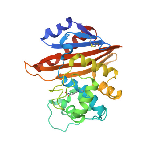
3LCE

Crystal Structure of Oxa-10 Beta-Lactamase Covalently Bound to Cyclobutanone Beta-Lactam Mimic


Experimental Data Snapshot

  • Method: X-RAY DIFFRACTION
  • Resolution: 2.00 Å
  • R-Value Free: 0.233 
  • R-Value Work: 0.178 
  • R-Value Observed: 0.181 

Starting Model: experimental
View more details

wwPDB Validation   3D Report Full Report


Ligand Structure Quality Assessment 


This is version 1.3 of the entry. See complete history


Literature

Cyclobutanone Analogues of beta-Lactams Revisited: Insights into Conformational Requirements for Inhibition of Serine- and Metallo-beta-Lactamases.

Johnson, J.W.Gretes, M.Goodfellow, V.J.Marrone, L.Heynen, M.L.Strynadka, N.C.Dmitrienko, G.I.

(2010) J Am Chem Soc 132: 2558-2560

  • DOI: https://doi.org/10.1021/ja9086374
  • Primary Citation of Related Structures:  
    3LCE

  • PubMed Abstract: 

    The most important mode of bacterial resistance to beta-lactam antibiotics is the expression of beta-lactamases. New cyclobutanone analogues of penams and penems have been prepared and evaluated for inhibition of class A, B, C, and D beta-lactamases. Inhibitors which favor conformations in which the C4 carboxylate is equatorial were found to be more potent than those in which the carboxylate is axial, and molecular modeling studies with enzyme-inhibitor complexes indicate that an equatorial orientation of the carboxylate is required for binding to beta-lactamases. An X-ray structure of OXA-10 complexed with a cyclobutanone confirms that a serine hemiketal is formed in the active site and that the inhibitor adopts the exo envelope. An unsaturated penem analogue was also found to enhance the potency of meropenem against carbapenem-resistant MBL-producing strains of Chryseobacterium meningosepticum and Stenotrophomonas maltophilia. These cyclobutanones represent the first type of reversible inhibitors to show moderate (low micromolar) inhibition of both serine- and metallo-beta-lactamases and should be considered for further development into practical inhibitors.


  • Organizational Affiliation

    Department of Chemistry, University of Waterloo, 200 University Avenue West, Waterloo, Ontario, Canada N2L 3G1.


Macromolecules
Find similar proteins by:  (by identity cutoff)  |  3D Structure
Entity ID: 1
MoleculeChains Sequence LengthOrganismDetailsImage
Beta-lactamase OXA-10
A, B, C, D
246Pseudomonas aeruginosaMutation(s): 0 
EC: 3.5.2.6
UniProt
Find proteins for P14489 (Pseudomonas aeruginosa)
Explore P14489 
Go to UniProtKB:  P14489
Entity Groups  
Sequence Clusters30% Identity50% Identity70% Identity90% Identity95% Identity100% Identity
UniProt GroupP14489
Sequence Annotations
Expand
  • Reference Sequence
Small Molecules
Ligands 3 Unique
IDChains Name / Formula / InChI Key2D Diagram3D Interactions
LCE
Query on LCE

Download Ideal Coordinates CCD File 
H [auth A],
M [auth B]
(1S,3S,4S,5S)-7,7-dichloro-3-methoxy-2-thiabicyclo[3.2.0]heptan-6-one-4-carboxylic acid
C8 H8 Cl2 O4 S
PELFTKQNZKVHIO-NXTKUWPGSA-N
PO4
Query on PO4

Download Ideal Coordinates CCD File 
N [auth C],
P [auth D]
PHOSPHATE ION
O4 P
NBIIXXVUZAFLBC-UHFFFAOYSA-K
GOL
Query on GOL

Download Ideal Coordinates CCD File 
E [auth A]
F [auth A]
G [auth A]
I [auth B]
J [auth B]
E [auth A],
F [auth A],
G [auth A],
I [auth B],
J [auth B],
K [auth B],
L [auth B],
O [auth C],
Q [auth D],
R [auth D],
S [auth D]
GLYCEROL
C3 H8 O3
PEDCQBHIVMGVHV-UHFFFAOYSA-N
Modified Residues  1 Unique
IDChains TypeFormula2D DiagramParent
KCX
Query on KCX
A, B, C, D
L-PEPTIDE LINKINGC7 H14 N2 O4LYS
Binding Affinity Annotations 
IDSourceBinding Affinity
LCE PDBBind:  3LCE IC50: 1.56e+5 (nM) from 1 assay(s)
Experimental Data & Validation

Experimental Data

  • Method: X-RAY DIFFRACTION
  • Resolution: 2.00 Å
  • R-Value Free: 0.233 
  • R-Value Work: 0.178 
  • R-Value Observed: 0.181 
  • Space Group: P 1 21 1
Unit Cell:
Length ( Å )Angle ( ˚ )
a = 65.7α = 90
b = 82.544β = 94.63
c = 102.36γ = 90
Software Package:
Software NamePurpose
MOSFLMdata reduction
SCALAdata scaling
REFMACrefinement
PDB_EXTRACTdata extraction
MAR345dtbdata collection
REFMACphasing

Structure Validation

View Full Validation Report


Ligand Structure Quality Assessment 


Entry History 

Deposition Data

Revision History  (Full details and data files)

  • Version 1.0: 2010-03-09
    Type: Initial release
  • Version 1.1: 2011-07-13
    Changes: Version format compliance
  • Version 1.2: 2023-09-06
    Changes: Data collection, Database references, Derived calculations, Refinement description
  • Version 1.3: 2023-11-22
    Changes: Data collection