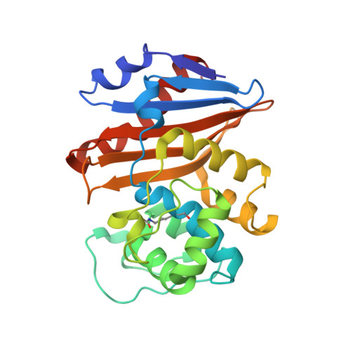
A focused fragment library targeting the antibiotic resistance enzyme - Oxacillinase-48: Synthesis, structural evaluation and inhibitor design.
Akhter, S., Lund, B.A., Ismael, A., Langer, M., Isaksson, J., Christopeit, T., Leiros, H.S., Bayer, A.(2018) Eur J Med Chem 145: 634-648
- PubMed: 29348071
- DOI: https://doi.org/10.1016/j.ejmech.2017.12.085
- Primary Citation of Related Structures:
5QA4, 5QA5, 5QA6, 5QA7, 5QA8, 5QA9, 5QAA, 5QAB, 5QAC, 5QAD, 5QAE, 5QAF, 5QAG, 5QAH, 5QAI, 5QAJ, 5QAK, 5QAL, 5QAM, 5QAN, 5QAO, 5QAP, 5QAQ, 5QAR, 5QAS, 5QAT, 5QAU, 5QAV, 5QAW, 5QAX, 5QAY, 5QAZ, 5QB0, 5QB1, 5QB2, 5QB3, 5QB4 - PubMed Abstract:
β-Lactam antibiotics are of utmost importance when treating bacterial infections in the medical community. However, currently their utility is threatened by the emergence and spread of β-lactam resistance. The most prevalent resistance mechanism to β-lactam antibiotics is expression of β-lactamase enzymes. One way to overcome resistance caused by β-lactamases, is the development of β-lactamase inhibitors and today several β-lactamase inhibitors e.g. avibactam, are approved in the clinic. Our focus is the oxacillinase-48 (OXA-48), an enzyme reported to spread rapidly across the world and commonly identified in Escherichia coli and Klebsiella pneumoniae. To guide inhibitor design, we used diversely substituted 3-aryl and 3-heteroaryl benzoic acids to probe the active site of OXA-48 for useful enzyme-inhibitor interactions. In the presented study, a focused fragment library containing 49 3-substituted benzoic acid derivatives were synthesised and biochemically characterized. Based on crystallographic data from 33 fragment-enzyme complexes, the fragments could be classified into R 1 or R 2 binders by their overall binding conformation in relation to the binding of the R 1 and R 2 side groups of imipenem. Moreover, binding interactions attractive for future inhibitor design were found and their usefulness explored by the rational design and evaluation of merged inhibitors from orthogonally binding fragments. The best inhibitors among the resulting 3,5-disubstituted benzoic acids showed inhibitory potential in the low micromolar range (IC 50 = 2.9 μM). For these inhibitors, the complex X-ray structures revealed non-covalent binding to Arg250, Arg214 and Tyr211 in the active site and the interactions observed with the mono-substituted fragments were also identified in the merged structures.
- Department of Chemistry, Faculty of Science and Technology, UiT- The Arctic University of Norway, N-9037 Tromsø, Norway.
Organizational Affiliation:


