6NBW

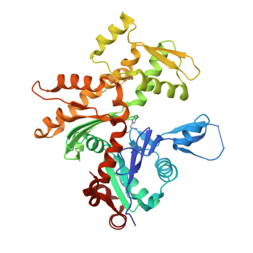
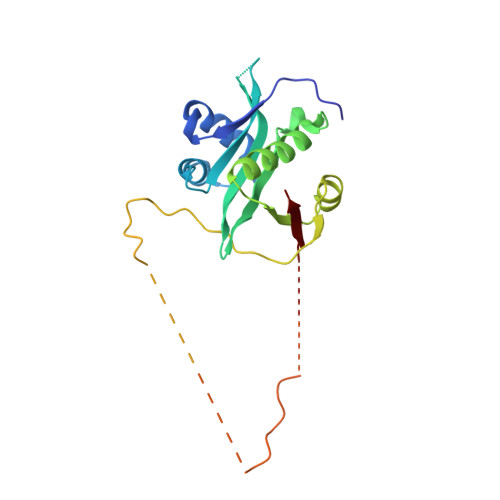
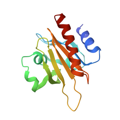
Ternary Complex of Beta/Gamma-Actin with Profilin and AnCoA-NAA80


Experimental Data Snapshot

  • Method: X-RAY DIFFRACTION
  • Resolution: 2.50 Å
  • R-Value Free: 0.209 
  • R-Value Work: 0.178 
  • R-Value Observed: 0.179 

Starting Model: experimental
View more details

wwPDB Validation   3D Report Full Report


Ligand Structure Quality Assessment 


This is version 1.3 of the entry. See complete history


Literature

Mechanism of actin N-terminal acetylation.

Rebowski, G.Boczkowska, M.Drazic, A.Ree, R.Goris, M.Arnesen, T.Dominguez, R.

(2020) Sci Adv 6: eaay8793-eaay8793

  • DOI: https://doi.org/10.1126/sciadv.aay8793
  • Primary Citation of Related Structures:  
    6NAS, 6NBE, 6NBW

  • PubMed Abstract: 

    About 80% of human proteins are amino-terminally acetylated (Nt-acetylated) by one of seven Nt-acetyltransferases (NATs). Actin, the most abundant protein in the cytoplasm, has its own dedicated NAT, NAA80, which acts posttranslationally and affects cytoskeleton assembly and cell motility. Here, we show that NAA80 does not associate with filamentous actin in cells, and its natural substrate is the monomeric actin-profilin complex, consistent with Nt-acetylation preceding polymerization. NAA80 Nt-acetylates actin-profilin much more efficiently than actin alone, suggesting that profilin acts as a chaperone for actin Nt-acetylation. We determined crystal structures of the NAA80-actin-profilin ternary complex, representing different actin isoforms and different states of the catalytic reaction and revealing the first structure of NAT-substrate complex at atomic resolution. The structural, biochemical, and cellular analysis of mutants shows how NAA80 has evolved to specifically recognize actin among all cellular proteins while targeting all six actin isoforms, which differ the most at the amino terminus.


  • Organizational Affiliation

    Department of Physiology, Perelman School of Medicine, University of Pennsylvania, Philadelphia, PA, USA.


Macromolecules
Find similar proteins by:  (by identity cutoff)  |  3D Structure
Entity ID: 1
MoleculeChains Sequence LengthOrganismDetailsImage
Actin, cytoplasmic 1374Homo sapiensMutation(s): 0 
EC: 3.6.4
UniProt & NIH Common Fund Data Resources
Find proteins for P60709 (Homo sapiens)
Explore P60709 
Go to UniProtKB:  P60709
PHAROS:  P60709
GTEx:  ENSG00000075624 
Entity Groups  
Sequence Clusters30% Identity50% Identity70% Identity90% Identity95% Identity100% Identity
UniProt GroupP60709
Sequence Annotations
Expand
  • Reference Sequence
Find similar proteins by:  (by identity cutoff)  |  3D Structure
Entity ID: 2
MoleculeChains Sequence LengthOrganismDetailsImage
N-alpha-acetyltransferase 80B [auth N]235Homo sapiensMutation(s): 0 
Gene Names: NAA80FUS2NAT6
EC: 2.3.1
UniProt & NIH Common Fund Data Resources
Find proteins for Q93015 (Homo sapiens)
Explore Q93015 
Go to UniProtKB:  Q93015
PHAROS:  Q93015
GTEx:  ENSG00000243477 
Entity Groups  
Sequence Clusters30% Identity50% Identity70% Identity90% Identity95% Identity100% Identity
UniProt GroupQ93015
Sequence Annotations
Expand
  • Reference Sequence
Find similar proteins by:  (by identity cutoff)  |  3D Structure
Entity ID: 3
MoleculeChains Sequence LengthOrganismDetailsImage
Profilin-1C [auth P]140Homo sapiensMutation(s): 0 
Gene Names: PFN1
UniProt & NIH Common Fund Data Resources
Find proteins for P07737 (Homo sapiens)
Explore P07737 
Go to UniProtKB:  P07737
PHAROS:  P07737
GTEx:  ENSG00000108518 
Entity Groups  
Sequence Clusters30% Identity50% Identity70% Identity90% Identity95% Identity100% Identity
UniProt GroupP07737
Sequence Annotations
Expand
  • Reference Sequence
Small Molecules
Ligands 6 Unique
IDChains Name / Formula / InChI Key2D Diagram3D Interactions
SOP
Query on SOP

Download Ideal Coordinates CCD File 
I [auth N][(2R,3S,4R,5R)-5-(6-AMINO-9H-PURIN-9-YL)-4-HYDROXY-3-(PHOSPHONOOXY)TETRAHYDROFURAN-2-YL]METHYL (3R)-3-HYDROXY-2,2-DIMETHYL-4-OXO-4-{[3-OXO-3-({2-[(2-OXOPROPYL)THIO]ETHYL}AMINO)PROPYL]AMINO}BUTYL DIHYDROGEN DIPHOSPHATE
C24 H40 N7 O17 P3 S
GAMKENBUYYJLCQ-NDZSKPAWSA-N
ATP
Query on ATP

Download Ideal Coordinates CCD File 
E [auth A]ADENOSINE-5'-TRIPHOSPHATE
C10 H16 N5 O13 P3
ZKHQWZAMYRWXGA-KQYNXXCUSA-N
LAB
Query on LAB

Download Ideal Coordinates CCD File 
F [auth A]LATRUNCULIN B
C20 H29 N O5 S
NSHPHXHGRHSMIK-JRIKCGFMSA-N
MES
Query on MES

Download Ideal Coordinates CCD File 
J [auth N]2-(N-MORPHOLINO)-ETHANESULFONIC ACID
C6 H13 N O4 S
SXGZJKUKBWWHRA-UHFFFAOYSA-N
GOL
Query on GOL

Download Ideal Coordinates CCD File 
G [auth A],
H [auth A]
GLYCEROL
C3 H8 O3
PEDCQBHIVMGVHV-UHFFFAOYSA-N
CA
Query on CA

Download Ideal Coordinates CCD File 
D [auth A]CALCIUM ION
Ca
BHPQYMZQTOCNFJ-UHFFFAOYSA-N
Modified Residues  1 Unique
IDChains TypeFormula2D DiagramParent
HIC
Query on HIC
A
L-PEPTIDE LINKINGC7 H11 N3 O2HIS
Experimental Data & Validation

Experimental Data

  • Method: X-RAY DIFFRACTION
  • Resolution: 2.50 Å
  • R-Value Free: 0.209 
  • R-Value Work: 0.178 
  • R-Value Observed: 0.179 
  • Space Group: C 2 2 21
Unit Cell:
Length ( Å )Angle ( ˚ )
a = 104.406α = 90
b = 115.594β = 90
c = 132.195γ = 90
Software Package:
Software NamePurpose
PHENIXrefinement
HKL-2000data reduction
HKL-2000data scaling
PHENIXphasing

Structure Validation

View Full Validation Report


Ligand Structure Quality Assessment 


Entry History & Funding Information

Deposition Data


Funding OrganizationLocationGrant Number
National Institutes of Health/National Institute of General Medical Sciences (NIH/NIGMS)United StatesGM073791

Revision History  (Full details and data files)

  • Version 1.0: 2020-01-29
    Type: Initial release
  • Version 1.1: 2020-08-12
    Changes: Database references, Derived calculations
  • Version 1.2: 2022-03-16
    Changes: Author supporting evidence, Database references
  • Version 1.3: 2023-10-11
    Changes: Data collection, Refinement description