6YP9 | pdb_00006yp9

Rabbit muscle actin in complex with ADF-H and ATP-ATTO-488


Experimental Data Snapshot

  • Method: X-RAY DIFFRACTION
  • Resolution: 2.56 Å
  • R-Value Free: 
    0.230 (Depositor), 0.230 (DCC) 
  • R-Value Work: 
    0.174 (Depositor), 0.170 (DCC) 
  • R-Value Observed: 
    0.177 (Depositor) 

Starting Model: experimental
View more details

wwPDB Validation   3D Report Full Report


Ligand Structure Quality Assessment 


This is version 1.2 of the entry. See complete history


Literature

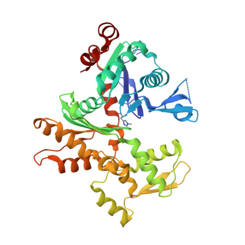
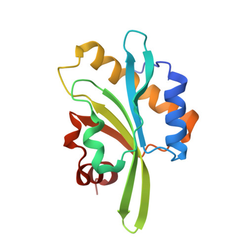
A functional family of fluorescent nucleotide analogues to investigate actin dynamics and energetics.

Colombo, J.Antkowiak, A.Kogan, K.Kotila, T.Elliott, J.Guillotin, A.Lappalainen, P.Michelot, A.

(2021) Nat Commun 12: 548-548

  • DOI: https://doi.org/10.1038/s41467-020-20827-4
  • Primary Citation of Related Structures:  
    6YP9

  • PubMed Abstract: 

    Actin polymerization provides force for vital processes of the eukaryotic cell, but our understanding of actin dynamics and energetics remains limited due to the lack of high-quality probes. Most current probes affect dynamics of actin or its interactions with actin-binding proteins (ABPs), and cannot track the bound nucleotide. Here, we identify a family of highly sensitive fluorescent nucleotide analogues structurally compatible with actin. We demonstrate that these fluorescent nucleotides bind to actin, maintain functional interactions with a number of essential ABPs, are hydrolyzed within actin filaments, and provide energy to power actin-based processes. These probes also enable monitoring actin assembly and nucleotide exchange with single-molecule microscopy and fluorescence anisotropy kinetics, therefore providing robust and highly versatile tools to study actin dynamics and functions of ABPs.


  • Organizational Affiliation
    • Aix Marseille Univ, CNRS, IBDM, Turing Centre for Living Systems, 13288, Marseille, France.

Macromolecules
Find similar proteins by:  (by identity cutoff)  |  3D Structure
Entity ID: 1
MoleculeChains Sequence LengthOrganismDetailsImage
Actin, alpha skeletal muscle375Oryctolagus cuniculusMutation(s): 0 
EC: 3.6.4
UniProt
Find proteins for P68135 (Oryctolagus cuniculus)
Explore P68135 
Go to UniProtKB:  P68135
Entity Groups  
Sequence Clusters30% Identity50% Identity70% Identity90% Identity95% Identity100% Identity
UniProt GroupP68135
Sequence Annotations
Expand
  • Reference Sequence
Find similar proteins by:  (by identity cutoff)  |  3D Structure
Entity ID: 2
MoleculeChains Sequence LengthOrganismDetailsImage
Twinfilin-1140Mus musculusMutation(s): 0 
Gene Names: Twf1Ptk9
UniProt & NIH Common Fund Data Resources
Find proteins for Q91YR1 (Mus musculus)
Explore Q91YR1 
Go to UniProtKB:  Q91YR1
IMPC:  MGI:1100520
Entity Groups  
Sequence Clusters30% Identity50% Identity70% Identity90% Identity95% Identity100% Identity
UniProt GroupQ91YR1
Sequence Annotations
Expand
  • Reference Sequence
Small Molecules
Modified Residues  1 Unique
IDChains TypeFormula2D DiagramParent
HIC
Query on HIC
A
L-PEPTIDE LINKINGC7 H11 N3 O2HIS
Experimental Data & Validation

Experimental Data

  • Method: X-RAY DIFFRACTION
  • Resolution: 2.56 Å
  • R-Value Free:  0.230 (Depositor), 0.230 (DCC) 
  • R-Value Work:  0.174 (Depositor), 0.170 (DCC) 
  • R-Value Observed: 0.177 (Depositor) 
Space Group: P 21 21 21
Unit Cell:
Length ( Å )Angle ( ˚ )
a = 50.552α = 90
b = 69.017β = 90
c = 147.948γ = 90
Software Package:
Software NamePurpose
BUSTERrefinement
XDSdata reduction
Aimlessdata scaling
PHASERphasing
autoPROCdata reduction

Structure Validation

View Full Validation Report


Ligand Structure Quality Assessment 


Entry History & Funding Information

Deposition Data


Funding OrganizationLocationGrant Number
Academy of FinlandFinland320161

Revision History  (Full details and data files)

  • Version 1.0: 2020-12-09
    Type: Initial release
  • Version 1.1: 2021-02-03
    Changes: Database references
  • Version 1.2: 2024-01-24
    Changes: Data collection, Database references, Refinement description