7LZ2

Structure of glutamate receptor-like channel GLR3.4 ligand-binding domain in complex with methionine


Experimental Data Snapshot

  • Method: X-RAY DIFFRACTION
  • Resolution: 1.50 Å
  • R-Value Free: 0.209 
  • R-Value Work: 0.185 
  • R-Value Observed: 0.186 

wwPDB Validation   3D Report Full Report


Ligand Structure Quality Assessment 


This is version 1.2 of the entry. See complete history


Literature

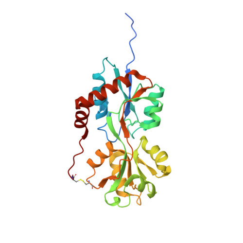
Structure of the Arabidopsis thaliana glutamate receptor-like channel GLR3.4.

Green, M.N.Gangwar, S.P.Michard, E.Simon, A.A.Portes, M.T.Barbosa-Caro, J.Wudick, M.M.Lizzio, M.A.Klykov, O.Yelshanskaya, M.V.Feijo, J.A.Sobolevsky, A.I.

(2021) Mol Cell 81: 3216

  • DOI: https://doi.org/10.1016/j.molcel.2021.05.025
  • Primary Citation of Related Structures:  
    7LZ0, 7LZ1, 7LZ2, 7LZH, 7LZI

  • PubMed Abstract: 

    Glutamate receptor-like channels (GLRs) play vital roles in various physiological processes in plants, such as wound response, stomatal aperture control, seed germination, root development, innate immune response, pollen tube growth, and morphogenesis. Despite the importance of GLRs, knowledge about their molecular organization is limited. Here we use X-ray crystallography and single-particle cryo-EM to solve structures of the Arabidopsis thaliana GLR3.4. Our structures reveal the tetrameric assembly of GLR3.4 subunits into a three-layer domain architecture, reminiscent of animal ionotropic glutamate receptors (iGluRs). However, the non-swapped arrangement between layers of GLR3.4 domains, binding of glutathione through S-glutathionylation of cysteine C205 inside the amino-terminal domain clamshell, unique symmetry, inter-domain interfaces, and ligand specificity distinguish GLR3.4 from representatives of the iGluR family and suggest distinct features of the GLR gating mechanism. Our work elaborates on the principles of GLR architecture and symmetry and provides a molecular template for deciphering GLR-dependent signaling mechanisms in plants.


  • Organizational Affiliation

    Department of Biochemistry and Molecular Biophysics, Columbia University, 650 West 168th Street, New York, NY 10032, USA; Training Program in Nutritional and Metabolic Biology, Institute of Human Nutrition, Columbia University Irving Medical Center, 630 West 168th Street, New York, NY 10032, USA.


Macromolecules
Find similar proteins by:  (by identity cutoff)  |  3D Structure
Entity ID: 1
MoleculeChains Sequence LengthOrganismDetailsImage
Glutamate receptor 3.4
A, B, C
268Arabidopsis thalianaMutation(s): 0 
Gene Names: GLR3.4GLR4GLUR3At1g05200YUP8H12.19
Membrane Entity: Yes 
UniProt
Find proteins for Q8GXJ4 (Arabidopsis thaliana)
Explore Q8GXJ4 
Go to UniProtKB:  Q8GXJ4
Entity Groups  
Sequence Clusters30% Identity50% Identity70% Identity90% Identity95% Identity100% Identity
UniProt GroupQ8GXJ4
Sequence Annotations
Expand
  • Reference Sequence
Small Molecules
Ligands 6 Unique
IDChains Name / Formula / InChI Key2D Diagram3D Interactions
MET (Subject of Investigation/LOI)
Query on MET

Download Ideal Coordinates CCD File 
D [auth A],
L [auth B],
P [auth C]
METHIONINE
C5 H11 N O2 S
FFEARJCKVFRZRR-BYPYZUCNSA-N
SO4
Query on SO4

Download Ideal Coordinates CCD File 
G [auth A],
N [auth B],
T [auth C],
U [auth C],
V [auth C]
SULFATE ION
O4 S
QAOWNCQODCNURD-UHFFFAOYSA-L
GOL
Query on GOL

Download Ideal Coordinates CCD File 
H [auth A],
I [auth A],
J [auth A],
O [auth B]
GLYCEROL
C3 H8 O3
PEDCQBHIVMGVHV-UHFFFAOYSA-N
BME
Query on BME

Download Ideal Coordinates CCD File 
K [auth A],
W [auth C]
BETA-MERCAPTOETHANOL
C2 H6 O S
DGVVWUTYPXICAM-UHFFFAOYSA-N
CL
Query on CL

Download Ideal Coordinates CCD File 
M [auth B],
Q [auth C],
R [auth C],
S [auth C]
CHLORIDE ION
Cl
VEXZGXHMUGYJMC-UHFFFAOYSA-M
NA
Query on NA

Download Ideal Coordinates CCD File 
E [auth A],
F [auth A]
SODIUM ION
Na
FKNQFGJONOIPTF-UHFFFAOYSA-N
Experimental Data & Validation

Experimental Data

  • Method: X-RAY DIFFRACTION
  • Resolution: 1.50 Å
  • R-Value Free: 0.209 
  • R-Value Work: 0.185 
  • R-Value Observed: 0.186 
  • Space Group: P 65 2 2
Unit Cell:
Length ( Å )Angle ( ˚ )
a = 74.581α = 90
b = 74.581β = 90
c = 508.887γ = 120
Software Package:
Software NamePurpose
XDSdata reduction
Aimlessdata scaling
PHASERphasing
REFMACrefinement
PDB_EXTRACTdata extraction

Structure Validation

View Full Validation Report


Ligand Structure Quality Assessment 


Entry History & Funding Information

Deposition Data


Funding OrganizationLocationGrant Number
National Institutes of Health/National Cancer Institute (NIH/NCI)United StatesR01 CA206573
National Institutes of Health/National Institute of Neurological Disorders and Stroke (NIH/NINDS)United StatesR01 NS083660
National Institutes of Health/National Institute of Neurological Disorders and Stroke (NIH/NINDS)United StatesR01 NS107253
National Science Foundation (NSF, United States)United States1818086

Revision History  (Full details and data files)

  • Version 1.0: 2021-07-28
    Type: Initial release
  • Version 1.1: 2021-08-18
    Changes: Database references
  • Version 1.2: 2023-10-18
    Changes: Data collection, Refinement description