Structural Insight into the Reaction Mechanism of Ketosynthase-Like Decarboxylase in a Loading Module of Modular Polyketide Synthases.
Chisuga, T., Nagai, A., Miyanaga, A., Goto, E., Kishikawa, K., Kudo, F., Eguchi, T.(2022) ACS Chem Biol 17: 198-206
- PubMed: 34985877
- DOI: https://doi.org/10.1021/acschembio.1c00856
- Primary Citation of Related Structures:
7VEE, 7VEF - PubMed Abstract:
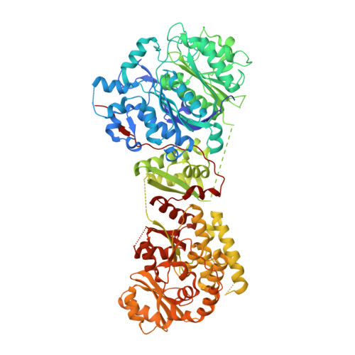
Ketosynthase-like decarboxylase (KS Q ) domains are widely distributed in the loading modules of modular polyketide synthases (PKSs) and are proposed to catalyze the decarboxylation of a malonyl or methylmalonyl unit for the construction of the PKS starter unit. KS Q domains have high sequence similarity to ketosynthase (KS) domains, which catalyze transacylation and decarboxylative condensation in polyketide and fatty acid biosynthesis, except that the catalytic Cys residue of KS domains is replaced by Gln in KS Q domains. Here, we present biochemical analyses of GfsA KS Q and CmiP4 KS Q , which are involved in the biosynthesis of FD-891 and cremimycin, respectively. In vitro analysis showed that these KS Q domains catalyze the decarboxylation of malonyl and methylmalonyl units. Furthermore, we determined the crystal structure of GfsA KS Q in complex with a malonyl thioester substrate analogue, which enabled identification of key amino acid residues involved in the decarboxylation reaction. The importance of these residues was confirmed by mutational analysis. On the basis of these findings, we propose a mechanism of the decarboxylation reaction catalyzed by GfsA KS Q . GfsA KS Q initiates decarboxylation by fixing the substrate in a suitable conformation for decarboxylation. The formation of enolate upon decarboxylation is assisted by two conserved threonine residues. Comparison of the structure of GfsA KS Q with those of KS domains suggests that the Gln residue in the active site of the KS Q domain mimics the acylated Cys residue in the active site of KS domains.
Organizational Affiliation:
Department of Chemistry, Tokyo Institute of Technology, 2-12-1 O̅okayama, Meguro-ku, Tokyo 152-8851, Japan.